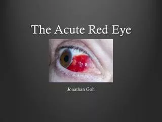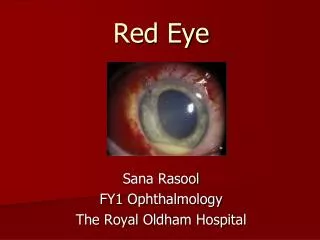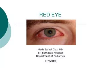
The Red Eye
WAOPS Spring Conference May 31, 2014 The Waters at Minocqua 8116 US 51 South Minocqua, WI Shiloh A. Simons, DO Ministry Medical Group Ophthalmology Stevens Point , WI. The Red Eye. History Symptoms: itching, discharge, irritation, pain, photophobia, blurred vision

The Red Eye
E N D
Presentation Transcript
WAOPS Spring Conference • May 31, 2014 • The Waters at Minocqua8116 US 51 SouthMinocqua, WI • Shiloh A. Simons, DO • Ministry Medical Group Ophthalmology • Stevens Point , WI The Red Eye
History Symptoms: itching, discharge, irritation, pain, photophobia, blurred vision Unilateral or bilateral presentation Character of discharge Recent exposure to an infected individual Trauma: mechanical, chemical, ultraviolet Contact lens wear: lens type, hygiene, and use regimen Systemic diseases (e.g., genitourinary discharge, dysuria, dysphagia, upper respiratory infection, skin and mucosal lesions) Allergy, asthma, eczema Use of topical and systemic medications Red Eye Workup
Physical Exam Measure Visual Acuity External Examination Pupil Exam, Motility Exam Slit-lamp examination Intraocular pressures Dilated Exam Red Eye Workup
External Exam Regional lymphadenopathy,particularly preauricular Skin: signs of rosacea, eczema, seborrhea Abnormalities of the eyelids: swelling, discoloration, malposition, laxity Conjunctiva: pattern of injection, subconjunctival hemorrhage, chemosis, cicatricial change Red Eye Workup
Slit-lamp Exam Eyelid margins: inflammation, vesicles Eyelashes: loss of lashes, trichiasis Lacrimalpuncta and tear film Conjunctiva: injection, papillae, follicles Cornea: Epithelial defects, punctatekeratopathy, dendrites, filaments, ulceration, subepithelial infiltrates Anterior chamber/iris: cells, flare, synechiae, transillumination defects Red Eye Workup
Diagnostic Testing Cultures: Bacterial, Viral, Chlamydial : Suspected cases of adult and in all cases of suspected neonatal conjunctivitis. Smears/Cytology: Smears for cytology and special stains (Gram, Giemsa) Blood Tests Biopsy: Conjunctival biopsy may be helpful in cases of conjunctivitis unresponsive to therapy. Red Eye Workup
Red Eye Diagnosis • Ocular Infections • Corneal Ulcers • Bacterial • Fungal • Acanthamoeba • OphthalmiaNeonatorum
Red Eye Diagnosis • Ocular Infections • Viral • Herpes Simplex • Herpes Zoster • Epidemic Keratoconjunctivitis • Hemorrhagic Conjunctivitis • PreseptalCellulitis • Orbital Cellulitis
Red Eye Diagnosis • Conjunctivitis • Allergic • Mechanical • Immune Mediated • Neoplasia
Red Eye Diagnosis • Trauma • Corneal Abrasion • Foreign Bodies • Subconjunctival Hemorrhage • Iritis • Chalazion • Nasolacrimal Duct Obstruction • Angle Closure Glaucoma
Ocular Infections • Corneal Ulcers • Bacterial • Fungal • Acanthamoeba • Viral
Ocular Infections • Bacterial • Staphylococci • 50% of the infections • Streptococci • Haemophilus • Pseudomonas • Serratia
Ocular Infections • Fungal • Candida • Gray white with feathery border • Fusarium • Outbreaks due to contact lens solution contaminant
Ocular Infections • Acanthamoeba • Contact lenses • Poor hygiene • Homemade solution • Swimming • Hot tubs • Extremely painful
Ocular Infections • Ophthalmia Neonatorum • Chemical • Neisseria Gonorrhoeae • Chlamydia Trachomatis • Staph, Strep, Gram Neg • Herpes Simplex Virus
Ocular Infections • Viral • Herpes Simplex Keratitis • Typical dendrite staining pattern • 90% exposure to virus by age 10
Ocular Infections • Herpes Zoster Ophthalmicus • Hutchinson’s Sign
Ocular Infections • Viral • Epidemic Keratoconjunctivitis • Adenovirus • Hemorrhagic Conjunctivitis • Coxsackie A
Ocular Infections • Preseptal Cellulitis • Tenderness, redness, swelling of lids • Minimal or no pain with eye movement • Dacryocystitis, sinusitis, trauma
Ocular Infections • Orbital Cellulitis • Pain on attempted eye movement • Proptosis, chemosis, fever • Admit to hospital • Trauma, sinusitis, surgery
Allergic Seasonal allergic conjunctivitis Vernal conjunctivitis Atopic conjunctivitis Giant papillary conjunctivitis (GPC), which also has a mechanical component Conjunctivitis
Conjunctivitis • Allergic • papillae • giant papillae
Mechanical Superior limbic keratoconjunctivitis (SLK) Contact-lens-related keratoconjunctivitis Floppy eyelid syndrome Pediculosispalpebrarum (Phthirus pubis) Medication-induced keratoconjunctivitis Conjunctivalchalasis Conjunctivitis
Mechanical Floppy eyelid syndrome Conjunctivitis
Immune-mediated Ocular mucous membrane pemphigoid (OMMP) Graft-versus-host disease (GVHD) Stevens-Johnson syndrome Conjunctivitis
Neoplastic Sebaceous (meibomian) carcinoma Ocular surface squamousneoplasia Melanoma Conjunctivitis
No entry into anterior chamber Decreased Vision Pain, usually improves with topical anesthesia Corneal Abrasion
Corneal Conjunctival Intraocular Orbital Foreign Bodies
Subconjunctival Hemorrhage • Typically not painful, not infection. • Often noticed by another or when looking in mirror.
Dull, aching, throbbing pain Photophobia Recurrent or initial, traumatic Iritis
Chalazion • Inflamed meibomian gland of eyelid • Usually sterile, granuloma
Nasal Lacrimal Duct Obstruction • Usually congenital and often clears by 1 year.
Acute Angle Closure Glaucoma • Eye/Orbit Pain, Headache • Blurred/Decreased Vision • Colored Halos • Nausea and Vomiting
Acute Angle Closure Glaucoma • Signs • Elevated intraocular pressure • Shallow anterior chamber • Corneal edema • Mid dilated pupil • Ciliary flush
Questions? shiloh.simons@ministryhealth.org (715) 342-7825 office (715) 340-2337 cell
References American Academy of Ophthalmology . Preferred Practice Patterns. San Francisco: American Academy of Ophthalmology, 2013. The Wills Eye Manual. 6th ed. Office and Emergency Room Diagnosis and Treatment of Eye Disease. Philadelphia: Lippincott Williams and Wilkins, 2012.