The Acute Red Eye
The Acute Red Eye. Jonathan Goh. Background . One of the most common eye complaints May present to GP, ED, or Optometrist Varied aetiologies (a lot!) Commonly self limiting / benign But – serious sight threatening pathology may present as an acute red eye. Initial approach. History

The Acute Red Eye
E N D
Presentation Transcript
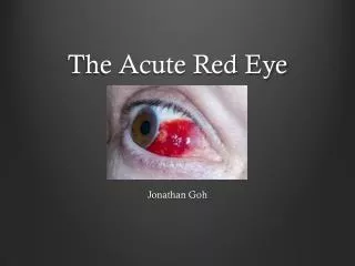
The Acute Red Eye Jonathan Goh
Background • One of the most common eye complaints • May present to GP, ED, or Optometrist • Varied aetiologies (a lot!) • Commonly self limiting / benign • But – serious sight threatening pathology may present as an acute red eye
Initial approach • History • Onset • Location – bilateral / unilateral / sectoral • Painful or painless – discomfort, gritty, foreign body sensation, itch, ache, sharp, pain on movement • Visual disturbance • Photosensitivity • Discharge – watery or purulent • Trauma to eye – e.g. hammering • ?contact lens • Anyone else with red eye • Recent travel • POHx and PMHx
Examination • Inspect whole patient • Visual Acuity + pin hole • Conjunctiva – bulbar and palpebral (evert lid) • Sclera • Cornea – clarity, fluorescein (abrasions, ulcers), sensation • Pupil – shape, reaction, accomodation • Eye movements – painful? Full?, diplopia? • Fundoscopy / slit lamp • Fluorescein • Tonometry • Lymph nodes - preauricular
Common Causes • Conjunctiva • Conjunctivitis • Bacterial • Viral • Allergic • Subconjhaemorrhage • Scleritis and episcleritis • Pterygium • Pingueculum • Cornea • Ulcer / abraision • Bacterial keratitis • Herpetic keratiis • Foreign body • Anterior chamber • Anterior uveitis/iritis/vitritis • Eye lids • Blepharitis • Chalazion / stye • Sub tarsal FB • Canaliculitis • Dacrocystitis • Marginal keratitis • Angle-closure glaucoma • Herpes Zoster ophthalmicus • Trauma • Preseptal and orbital cellulitis
Case 1 • 28 YO male • Previously well • 2 day history of red eyes, grittiness and mucopurulent discharge • Difficulty opening eyes on waking • Vision clears on blinking
Conjunctivitis • Bacterial • Viral • Allergic • Gonoccocal / Chlamydial
Conjunctivitis - Bacterial • Usually bilateral (within 48 hrs) • 70% Gram+ve: Streptococcus pneumoniae, Staphylococcus aureus • 30% Gram-ve: Haemophilusinfluenzae, Morxellacatarrhalis • Symptoms: Grittiness / burning, mucopurulent discharge, matting of eye lids, crusting, NO photophobia, NO visual disturbance • Signs: Crusty/purulent lids, conjunctival hyperaemia, mild papillary reaction, oedematous conjunctiva/lids,diffuse injection of conjunctiva (tends to be worse in fornices) • NO corneal or anterior chamber involvement • Treatment: Hygiene, topical antibiotics for 5 days (e.g. chloramphenicol)
Conjunctivitis - Viral • Acute onset • Uni or bilateral • Usually adenovirus type 3, 4,or 7 • History of URTI, may be epidemic • May develop late keratitis • Symptoms: Grittiness, watery/serous discharge, NO visual disturbance • Signs: Watery, discharge, Preauricular LN, diffuse conj injection, eye lid oedema, follicles • Treatment: Supportive, hygeine, eye lubricants, may take weeks to resolve
Conjunctivitis - Allergic • IgE mediated • Tends to be seasonal • Bilateral • Symptoms: itch, +/- watery discharge, NO visual disturbance • FHx of atopy • Signs: diffuse conj injection bilaterally, papillae, chemosis, mild eyelid swelling • Treatment: avoid allergen, cold compresses, topical antihistamines, mast cell stabiliser, NSAIDs, vasoconstrictor
Conjunctivitis – Chlamydial and Gonococcal • Sexually active – genitals>hand>eye • (can also occur in new born via birth canal) • Chlamydial: subacute, FB sensation, purulent discharge, preauricular LN • Gonococcal: Hyperacute presentation with purulent discharge +++, chemosis, papillary reaction, preauricular LN, May lead to infection keratitis • Swab – N gonorrhoea: microscopy G-vediplococci, cultures • Treatment: refer to ophthalmologist, systemic antibiotics • Workup for STIs
Case 2 • 70 YO F • Noticed that part of the white of her eye became bright red after a bout of coughing. • No pain, no visual disturbance, no discharge. • PHx: AF (warfarin), T2DM, COPD, HTN
SubconjunctivalHaemorrhage • Due to bleeding of conjunctival or episcleral vessel • Spontaneous, trauma, systemic illness, anticoagulation, unilateral • Hx of anticoagulants/platelets, bleeding disorder, trauma/rubbing, coughing/vomiting • Symptoms – red eye, no visual disturbance or pain or discharge • Ensure no penetrating injury • Check BP, INR (warfarin), lubricate, reassure
Case 3 • 24 YO male apprentice welder presents at 8pm • Previously well • Sudden onset foreign body sensation, photophobia, tearing, mild conjunctival redness, some visual deterioration.
Ultraviolet keratitis / flash burn • Tends to occur 8-12 hours after exposure • UV damages corneal epithelium • Symptoms: Foreign body sensation, tearing, blurring of vision, photophobia • Signs: Superficial punctate keratitis (stains with fluorescein), conjunctival injection, chemosis, belpharospasm • Treatment: Epithelium usually recovers in 1-3 days, lubricants, analgesia, mydriatics
Case 4 • 35YO male • Previously well • Poked in right eye • Immediately complains of FB sensation, photophobia, tearing, red eye, decreased vision.
Corneal abrasion • Corneal epithelial defect • Commonly due to trauma • Symptoms: pain, FB sensation, photophobia, tearing, conjunctival injection • Signs: corneal epithelial defect, stains with fluorescein, FB under eyelid • Treatment: topical antibiotics, lubricants, analgesia
Case 5 • 5 YO boy • Previously well • Reaching up to grab something from a shelf in laundry, accidentally spills ammonia on face. • Comes in crying, painful red eyes, and decreased vision.
Corneal chemical burn • Ophthalmic emergency • Acid or alkali • Alkali penetrate further. Acids coagulate protein forming a protective barrier • Causes necrosis of conjunctival and corneal epithelium and stroma possibly leading to perforation. • Can lead to corneal opacification, vascularisation, symblepharon • Treatment: COPIOUS IRRIGATION, sweep fornices, urgent referral to ophthalmologist, analgesia
Corneal Ulcer • Destruction of epithlium and stroma due to an infection • Risk factors: contact lens, trauma, ocular surface disease, immunosupression • Bacterial • Often Hx of contact lens use • Epithelial defect + opacified base • Bacterial Staph epidermidis, Strep pneumoniae, Strep pyogenes, Haemophilusinfluenza, Morazellacatarrhalis, Neisseria spp. • Symptoms: pain, watering/discharge, blurred vision, photophobia, discharge • Signs: Corneal ulcer, corneal oedema, hypopyon, chemosis, hypopyon • Treatment: Urgent referral to ophthalmologist, never patch, cultures, topical antibiotics. • Fungal • Aspergillus, Candida, or Fusarium • Satalite infiltrates common, feathery edges • Hx of trauma with organic material
Corneal Ulcer • Viral • Herpes Simplex Virus • Usually due to reactivation of Type 1 (can be Type 2) • Involvement of CNV1 • Hx of stress / immunosupression • Symptoms: photophobia, tearing, pain • Signs: Dendritic ulcer with terminal bulbs, Reduced corneal sensation, Hutchinson’s sign • Treatment: urgent referral to ophthalmologist • Usually topical antiviral treatment + mydriatic
Case 6 • 30 YO female • Previously well • Presents with unilateral red eye with mild pain. States she had a similar episode a few months ago which resolved by itself.
Episcleritis and scleritis • Episcleritis: inflammation of the episclera (thin membrane covering sclera) • Causes: Idiopathic, associated with vascular/connective tissue disorders • Rapid onset, grittiness, dull headache, +/- watery discharge, NO visual disturbance • Focal areas affected – radial configuration of vessels • Usually self limiting, may be recurrent • Scleritis: inflammation of sclera • Infectious, autoimmune mediated • May have visual disturbance • Scleral oedema/discoloured, congestion of scleral plexus, irregular blood vessels • Nodular, diffuse, necrotizing • Anterior, posterior • Treatment: URGENT REFERAL to ophthalmologist
Acute angle-closure glaucoma • Due to iris blocking trabecular meshwork outflow tract resulting in raised IOP • Damages optic nerve head • Worsened by mydriasis– pupil dilation • Symptoms: severe ocular pain, blurred vision, halos, headache, nausea/vomiting, abdominal pain. • Signs: diffuse injection, corneal oedema (hazy), pupil fixed irregular and mid dilated, raised IOP, ciliary injection • Treatment: urgent referral to ophthalmologist, aim is to reduce IOP • Acetazolamide, glycerol, mannitol, topical timolol, prednisolone acetate, pilocarpine (miosis, opens TM), peripheral iridotomy
Uveitis • Inflammation of the iris, ciliary body or choroid. • Anterior (iris and ciliary body) • 50-70% idiopathic, associated with systemic diseases, infective (TB, syphilis, leprosy, HSV, HZV, HIV, fungal) • Sudden onset, red painful eye, tearing, visual disturbance, photophobia • Perilimbal injection, flare and cells in AC, keratic precipitates, hypopyon, pupil sluggish • Treatment: Urgent ophthalmology referral, mydriatics, analgesia, steroids (after consult with ophthal) • May need work up for vascular/inflammatory disorders • Consequences: cataracts, glaucoma, retinal detachments, band keratopathy
Pitfalls • Beware the “unilateral bacterial conjunctivitis” • Always check visual acuity • Don’t patch corneal ulcers • Call for help early