The Acute Red Eye
240 likes | 891 Vues
The Acute Red Eye. Sijie Heng President, Ophthalmology University Society of Glasgow (OpUS-G). An acute red eye. Scenario

The Acute Red Eye
E N D
Presentation Transcript
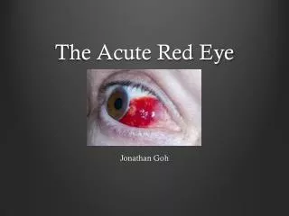
The Acute Red Eye Sijie Heng President, Ophthalmology University Society of Glasgow (OpUS-G)
An acute red eye • Scenario • While on an attachment in General Practice you are called to Mrs Jones who is a 60-year old woman who complains of the sudden development of a painful red right eye. She had had no previous episodes of a similar nature.
Pathophysiology • Dilatation of ocular surface vessels due to: • Infection • Inflammation • Allergy • Increased intraocular pressure
Intraorbital infections • Orbital cellulitis • Proptosis, pain ( on EM), EM, swollen (tender) eyelids • Systemic signs – fever, malaise • Usually history of long-standing sinusitis • Endophthalmitis • Pain (±), VA, hypopyon • History of ocular surgery/trauma, immunosuppression, infection
Orbital Cellulitis Pictures from emedicine.medscape.com
Acute angle-closure glaucoma • Pain, N & V, VA, haloes • Oval pupil, cloudy cornea, loss of red reflex, ‘rock-hard’ eye • Hypermetropes, dilating eye drops, FHx • Can be secondary e.g. iritis
Acute angle closure glaucoma Picture from University of Michigan Kellogg Eye Center, available from aao.org
Keratitis/Corneal Ulcers • Pain (!!), FB sensation, blurring, ± discharge, photophobia • Corneal opacities, hypopyon, fluorescein stain + • Contact lens, trauma/FB, lid disease • Beware: FB, HSV, HZV (shingles), neurotrophic
Corneal Ulcer with Hypopyon Picture from brooksidepress.org
Iritis • Also anterior uveitis, iridocyclitis • Pain (aching, on reading), photophobia (++), lacrimation, blurring • Miosis, corneal precipitates, AC ‘flare’ • PMHx: • Ocular: HSV, HZV, trauma • Systemic: HLA-B27 syndromes, JIA, sarcoid, syphilis
Iritis with keratic precipitates (KPs) Picture from mrcophth.com
Scleritis • Pain (++, on EM, night), blurring, photophobia, lacrimation • Tenderness (++), segmental/diffuse, deep engorged vessels (phenylephrine -), ± nodules • Hx: HZO, RA, SLE
Scleritis Picture from gp-training.net
Trauma/Foreign body • vision, pain, bright red blood (SCH), irritation, profuse watering • Bruising, SCH, hyphaema, corneal defects • Mechanism: superficial, blunt, perforating • Beware: children, intraocular involvement
Hyphaema & subconjunctival haemorrhage Picture from casereports.bmj.com
Conjunctivitis • Itch, burning, ‘grittiness’, vision preserved • 3 broad varieties: • Bacterial: purulent discharge • Viral: follicles, pre-auricular lymphadenopathy • Allergic: Hx of atopy, ‘cobblestone’ • Beware: cornea,neonates, trachoma (chlamydia), FB, systemic conditions (SJS, Erythema multiforme, Kawasaki’s, Reiter’s etc.)
Allergic conjunctivtis with giant papillae Picture from eyequestions.com
A systematic approach • A good history • General inspection • Functional: • Best-corrected visual acuity, visual fields • Pupil size + reactivity, EOM • Structural: • Periorbital tissues: lesions, swelling/proptosis • Conjunctivae (pull, evert) • Sclerae: segmental, nodules • Corneas: defects, opacities • Anterior chamber, red reflex, fundus • Elsewhere – rashes, joints, lymphadenopathy
Red Flags • Severe pain • Systemic features – fever, malaise etc. • Nausea and vomiting, headaches • Decreased visual acuity • Corneal lesions • Zoster skin rash • Penetrating trauma • If you don’t have a clue, but concerned
Summary • Red eye is common • Majority conjunctivitis • Severe pain, visual loss, photophobia referral • >1 condition(s) simultaneously • If in doubt, seek a senior/ ophthalmology opinion
Thank you for your attention • Questions? • Feedback please! • Visit opusg.webs.com
Bibliography Olver J, Cassidy L. Ophthalmology at a Glance. Oxford: Blackwell; 2009. Batterbury M, Bowling B, Murphy C. Ophthalmology: An illustrated colour text. 3rd edition. UK: Elsevier; 2009. PT Khaw, Shah P, Elkington AR. ABC of Eyes. 4th edition. London: BMJ; 2004. Merck’s Manual on Red eye by Kathryn Colby (updated April 2009, accessed October 2011) Available from: http://www.merckmanuals.com/professional/eye_disorders/symptoms_of_ophthalmologic_disorders/red_eye.html