Download
1 / 27
290 likes | 537 Vues
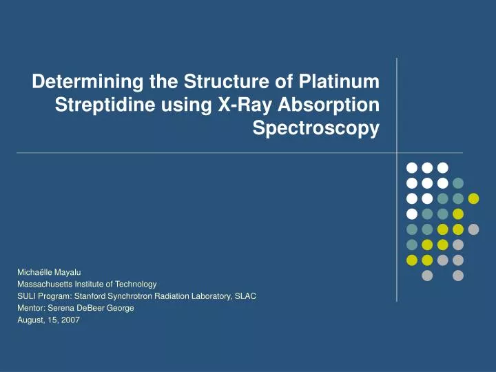
Determining the Structure of Platinum Streptidine using X-Ray Absorption Spectroscopy. Micha ë lle Mayalu Massachusetts Institute of Technology SULI Program: Stanford Synchrotron Radiation Laboratory, SLAC Mentor: Serena DeBeer George August, 15, 2007. Outline. Platinum Anti-Cancer Agents

E N D
Determining the Structure of Platinum Streptidine using X-Ray Absorption Spectroscopy Michaëlle Mayalu Massachusetts Institute of Technology SULI Program: Stanford Synchrotron Radiation Laboratory, SLAC Mentor: Serena DeBeer George August, 15, 2007
Outline • Platinum Anti-Cancer Agents • X-Ray Absorption Spectroscopy • Data Analysis • Conclusion
Platinum Anti-Cancer Agents Background Platinum Streptidine
Background • Platinum complexes have been discovered to stop cell division • This has useful applications in treating cancer and considerable advances have been made However….. • mechanisms are that lead to the drug being taken up by the cell membrane and integrated into the DNA are still unknown • the drug is successful in treating some types of cancers but ineffectual treating other types • platinum drugs such as cisplatin has been found to be very toxic causing nephrotoxity, neuropathy, ototoxicity, hematological toxicity, neuropathology and seizures. • Consequently, the search for improved platinum drugs that treat a wider range of cancers and display fewer toxic side effects still continues.
Platinum Streptidine • Because the structure and coordination of the drug (especially in solution) is essential to understanding how the drug interacts with molecules in the body, X-ray absorption spectroscopy (XAS) is a powerful tool for determining the local structure of this newly developed drug. • main questions to hopefully be answered by analysis of XAS data measured with XAS are: • Is it the Pt coordinated to the Streptidine? • And if this is the case through which atoms • Putative structures (obtained from elemental analysis and nuclear magnetic resonance) • Streptidine N. Aliaga-Alcalde and Jan Reedijk
X-Ray Absorption Spectroscopy What Is It Measurements Applications
What is it? Auger electron Emitted photo-electron continuum X-ray Fluorescence 2p Direct Absorption (Transmittance) 2s h h fluorescent photon 1s George DeBeer, S.
What is it? Pt-L3 Edge 11550 eV www.chem.ucalgary/groups/farideh/xas.pdf
Experimental Hutch X-ray Source Ta Slits Sample Foil I0 I1 I2 Double Crystal Monochromator Mono slits Detector Measurements George DeBeer, S.
Measurements: Transmittance • When a beam of monochromatic X-rays goes through matter, it loses its intensity due to interaction with the atoms in the material. The intensity drops exponentially with distance if the material is homogeneous, and after transmission the intensity is: I0=Ie-μt • Where: • I incident X-ray intensity • I0 transmitted X-ray intensity • µ absorption coefficient • t is the thickness of the sample • Absorption can therefore be measured as: A= µt=ln(I0/I) www.ssrl.slac.stanford.edu/dichroism/xas
Transmittance t A = μt = ln (I0/I1) μ ,absorption coefficient t sample thickness Ion chamber Ion chamber X-rays I0 I Sample + X-rays - Ionizing radiation (i.e. X-rays) creates ion pairs in the gas and the sweeping voltage results in a current flow. George DeBeer, S.
Fluorescence detector Soller Slits filter X-ray sample Measurements: Fluorescence George DeBeer, S.
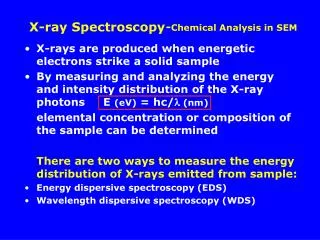
Applications X-Ray Absorption Spectrum (edge + EXAFS) EXAFS (extended x-ray absorption fine structure) Pre-edge and Edge (XANES) Absorption Coefficient (mu) Energy XAS or XAFS George DeBeer, S.
Applications Extended X-Ray absorption fine structure constructive interference results in a maximum destructive interference results in a minimum George DeBeer, S.
Data Analysis Processing Data Analyzing Results Fitting Data
Getting the EXAFS 1.4 1.2 Raw data: This is the way that the XAS transmission mode spectrum looks, right off the beam line. 1.0 Raw 0.8 0.6 0.4 6800 7000 7200 7400 7600 7800 8000 Pre-edge subtraction: A procedure performed to subtract the total absorption from the absorption of the edge in interest. 1.2 1.0 Raw 0.8 0.6 0.4 6800 7000 7200 7400 7600 7800 8000 eV 1.5 Spline: A method for removing the atomic background from the absorption curve (i.e. the absorption due to the photoabsorber alone, with out any neighboring atoms) . 1.0 Normal 0.5 0.0 6800 7000 7200 7400 7600 7800 8000 Processing Data eV 1.4 George DeBeer, S.
15.0 10.0 FT Magnitude 5.0 0.0 0 1 2 3 4 5 6 R (Å) Processing Data Getting Information from EXAFS Data A Fourier transform allows you to visualize the radial distribution of atoms. Note: k is the photoelectron wave number. k= (2m(E-E0)/ћ2)1/2 EXAFS data are k-weighted to enhance oscillations at high-k. EXAFS data is really a sum of sine waves. The goal of fitting data is to deconvolute the total signal into its components. George DeBeer, S.
Analyzing Results Pt-Std Edges Solid and Solution • As can be seen, the edges of the solid and solution data begin at different energies • Because the solid begins at a lower energy, it is more reduced • This suggests that the solid structure is surrounded by heavier atoms solid soln
Analyzing Results K3 EXAFS and Corresponding Fourier Transforms • amplitude of the EXAFS slightly decreases from solid to soln • this indicate a decrease in coordination number or may also be indicative of lighter atoms present in solution. • intensity of FT greatly decreases from solid to solution. • Similar to the decrease in EXAFS amplitude, the decrease in peak intensity indicates a decrease in coordination or lighter atoms ligated to the Platinum in solution.
Fitting Data Initial Model Calculation of initial distance & disorder parameters. Optimization of distance & disorder parameters. Are fit parameters reasonable & is fit quality good? Compare with other good fits to determine best fit. No Yes George DeBeer, S.
Solid Pt-Std Best Fit: 4 Pt-Cl at 2.30 Å and 1 Pt-N at 2.02 Å 4 Pt-Cl at 2.30 Å Normalized error 0.567 F/(No. pts) Normalized error: 0.547F/(No. pts) Cl Cl Pt Cl Cl N Comparison of K2PtCl4and C8H18N6O7Cl4Pt
Solution Pt-Std 4 Pt-Cl/S at 2.30 Å 1 Pt-N at 2.03 Å Normalized error: 0.359F/(No. pts)
Solution Pt-Std Best Fit: 3 Pt-Cl/S at 2.30 Å and 2 Pt-N at 2.08 Å Cl/S Normalized error: 0.363 F/(No. pts) Cl/S Cl/S Pt N N
Conclusion • It can be concluded that Platinum is in fact coordinated to the streptidine through a bond with nitrogen. • In the solid… • the streptidine is coordinated to the Pt by one nitrogen at 2.02 Å • In the solution… • the streptidine could be coordinated to 1-2 nitrogens, although with 2 nitrogens at 2.08 Å is more likely. • This is shown by analysis and fitting of the EXAFS data of the solid and solution Pt-Std and comparing the edges and EXAFS of the Pt-Std solid and solution • Future work should include the determination of how many chlorines and sulfurs are ligated in the solution structure.
Acknowledgments • Special Thanks to: • My Mentor Serena Debeer George • U. S. Department of Energy, Office of Science for giving me the opportunity to participate in the SULI
References • Serena DeBeer George, Introduction to Extended X-ray Absorption Fine Structure (EXAFS) Spectroscopy and its Applications (Power Point) • XAS Short Course for Structural Molecular Biology Applications • www.chem.ucalgary/groups/farideh/xas.pdf • www.ssrl.slac.stanford.edu/dichroism/xas • N. Aliaga-Alcalde and Jan Reedijk, University of Leiden, the Netherlands, unpublished results,Pt-Std (Platinum-Streptidine complexes)