Download
1 / 14
140 likes | 187 Vues
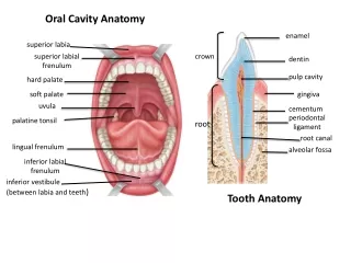
BASIC ORAL ANATOMY. Oral Cavity ( mouth). The entire oral cavity is lined with mucous membrane tissue. The oral cavity consists of the following two areas: The vestibule is the space between the teeth and the inner mucosal lining of the lips and checks.

E N D
Oral Cavity (mouth) • The entire oral cavity is lined with mucous membrane tissue. The oral cavity consists of the following two areas: • The vestibule is the space between the teeth and the inner mucosal lining of the lips and checks. • The oral cavity proper is the space contained within the upper and lower dental arches.
The Dentitions • The term dentition is used to describe the natural teeth in the jawbones. • Primary dentition is the first set of 20 primary teeth. Also referred to as “baby teeth” or “deciduous teeth” • Permanent dentition refers to the 32 secondary or “adult” teeth. • Mixed dentitionoccurs when both primary and permanent teeth are present, usually between the ages of 6 to 12.
Dental arches • The maxillary arch (upper arch), actually part of the skull, is fixed and not capable of movement. The teeth in the upper arch are set in the maxilla, the maxillary bone. • The mandibular arch (lower arch) is capable of movement through the action of the temporomandibular joint. The mandible, the mandibular bone supports the teeth in the lower arch.
Quadrants • An imaginary midline divides each arch into a left half and a right half. When the maxillary and mandibular arches are each divided into halves the resulting four sections are called quadrants, as follows: • Maxillary right quadrant • Maxillary left quadrant • Mandibular right quadrant • Mandibular left quadrant
Directions of the Oral Cavity • Anterior means toward the front of the mouth. • Posterior means toward the back of the mouth. • Medial means toward the middle or toward the middle of the arch. • Lateral means toward the side or toward the outside of the mouth.
Mesial means toward the mid-line of the dental arch. • Distal means away from the mid-line of the dental arch.
Eruption & Exfoliation • Eruption is the movement of the tooth through the surrounding tissues so that more of the tooth becomes visible in the mouth. • Exfoliation is the process by which the roots of the baby tooth are resorbed and dissolved until so little root remains that the baby tooth falls out.
Occlusion • Occlusion is the relationship of the mandibular and maxillary teeth when closed or during excursive movements of the mandible; when the teeth of the mandibular arch come into contact with the teeth of the maxillary arch in any functional relationship.
Types of Teeth • The functions of teeth vary, depending on their individual shape and size and their location in the jaws. The three basic food processing functions of the teeth are cutting, holding or grasping, and grinding. • Incisors are single-rooted teeth with a relatively sharp thin edge referred to as the incisal edge. Located in the front of the mouth, they are designed to cut food without the application of heavy forces. Central (front teeth) and lateral (distal to the centrals) teeth are incisors.
Canines, also known as cuspids, are located at the corner of the arch. They are designed for cutting and tearing foods, which require the application of force. • Premolars are a cross between canines and molars. An older term for premolar is bicuspid. The pointed cusps hold and grind the food. They have a broader surface for chewing food. There are two sets of premolars in the permanent dentition and NO premolars in the primary dentition.
Molars are much larger than premolars. The molars have more cusps than other teeth that are used to chew or grind up food. There are two sets of molars in the primary dentition and three sets of molars in the permanent dentition.
In each quadrant there are five permanent teeth (central, lateral, canine, & premolars) that succeed or take the place of the five primary teeth (central, lateral, canine, & molars), they are called succedaneous teeth. Three permanent molars do not succeed primary teeth in each quadrant; therefore they are nonsuccedaneous teeth.
Dentition Occlusion Maxilla - maxillary Mandible –mandibular Midline Quadrant Anterior Posterior Medial Lateral (direction) Permanent – adult Oral cavity Incisor Central Lateral Canine - cuspid Premolar – bicuspid Molar Succedaneous Eruption Exfoliation Primary – deciduous – baby Vestibule Keywords