Role of VEGF C(-460)T Polymorphism in Renal Cell Carcinoma Development
10 likes | 92 Vues
Investigating how SNP in VEGF promoter affects RCC development, using a novel QPCR detection format for real-time PCR. Results show reliable discrimination between SNP variants, offering potential for risk assessment.

Role of VEGF C(-460)T Polymorphism in Renal Cell Carcinoma Development
E N D
Presentation Transcript
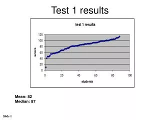
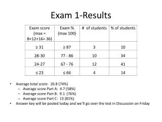
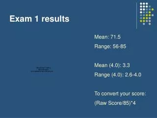
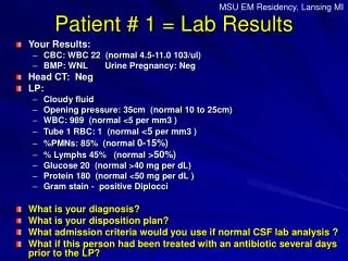
Role of VEGF C(-460)T single nucleotide polymorphism in the development of renal cell carcinoma S. Füssel1, S. Schneider1, A. Lohse-Fischer1, S. Tomasetti1, T. Köhler2, A. Rost2, A. Meye1, M.P. Wirth1 1Department of Urology, Medical Faculty, Technical University of Dresden & 2AJ Roboscreen Leipzig granted by technology support with financial sources of the European Regional Development Fund and the State of Saxony heterozygous homozygous mutant homozygous wild-type Q Q Q Q Q Q R1 R1 R1 5` 5` 5` 3` 5` 3` R2 R2 R2 5` 5` 5` 5` 5` 5` 5` 5` 3` 3` 3` 3` Fig. 1 TRIPLEHYB probe format: basic principle R1 = FAM R2 = ROX Q = BHQ1 / BHQ2 TT Du-145 CC patient CT LNCaP TT Du-145 CC patient CT LNCaP D1 – D4: positions for the attachment of different dyes D1 fluorescent dyeD2 quencher Fig. 4 Optimization of the SNP-detection assay FAM channel (for T-variant = mut)ROX channel (for C-variant = wt) Fig. 2 C(-460)T polymorphism in the VEGF promoter as model system Q = BHQ1 or BHQ2 R2 = ROX • plasmids and DNA from cell lines used as standards and controls for all variants • concordance with sequencing results (for 30 patient samples) 3-step PCR (45 cycles; LC480): 95°C/15s; 45°C/1s; 59°C/40s; primers: 0.5µM each; probes: 0.3µM ROX-up/0.4µM FAM-up + 1.2µM do; Universal Master Mix + add. 5mM MgCl2 (ABI); template: 10 / 20 / 50 / 100ng DNA • SNP at position 3 of the up-stream probe (acting as hydrolization probe) • perfect match binding to template amplification signal • no perfect match no binding to template no signal • labeling with fluorescent dyes (FAM, ROX) and quenchers (BHQ1+2) Fig. 3 Genotyping of the VEGF-SNP C(-460)T in a single reaction A fluorescence signal is only expected in case of perfect match: homozygous mut & heterozygous wt/mut positive in FAM channel homozygous wt & heterozygous wt/mut positive in ROX channel • Objectives • single nucleotide polymorphisms (SNPs) are frequently associated with the onset or progression of diseases including cancers • SNPs in the promoter region of the vascular endothelial growth factor (VEGF) are reported to influenceVEGF expression which is increased in several tumor types including renal cell carcinoma (RCC) • analysis of SNP distribution supports risk assessment and prediction of cancer development • determination of SNP variants by sequencing, array analysis or quantitative PCR (QPCR) • QPCR approaches include melting curve analyses or the use of SNP-specific probes • aim of the study: • establishment of a novel QPCR detection format for real-time PCR suitable for SNP detection • improved or same sensitivity, specificity, flexibility, and robustness compared to available formats • no need for external licenses (interest of the cooperating company AJ Roboscreen) • development of the TRIPLEHYB probe format & its validation in a clinico-experimental application • Results 1 • comparison of 5 systems containing the SNP at postion 1 to 5 of the up-stream probe systems 2-5 work well, system 3 offers most stable & reliable discrimination (Fig.4) • concordance between sequencing results & QPCR measurements based on system 3 • same results with patient DNA from tissue, whole blood & leukocytes • TRIPLEHYB as simple probe format for SNP determination fast and simple • Material & Methods • selection of the SNP C/T at postion -460 of the VEGF promoter as model system since it is supposed to alter VEGF expression analysis of this SNP in patients with clear-cell RCC • C corresponds to wild-type (wt) & T to mutant (mut) • design of a probe system based on the TRIPLEHYB format (Fig.1) consisting of: • the same forward & reverse primers used for both SNP variants • two up-stream probes each specific for one SNP variant • one down-stream probe matching to both up-stream probes • one half of the up-stream probe is complementary to one half of the down-stream probe forming a stem structure, the other halfs of the probes are complementary to the template (Fig. 1) • labeling with fluorescent dyes (D1) and quenchers (D2) specific combination for each SNP variant: ROX + BHQ2 for wt and FAM + BHQ1 for mut (Fig.2) • fluorescence signal only expected in case of perfect match between up-stream probe and template • parallel detection of amplification signals in both channels homozygous wt & heterozygous wt/mut positive in ROX channel, homozygous mut & heterozygous wt/mut positive in FAM channel (Fig.3) • design and testing of different probe systems containing the nucleotide substitution at postions 1-5 of the up-stream probe reliable discrimination of both SNP variants possible? • testing on cloned model plasmids for each SNP variant using the LightCylcer 480 (Roche) and the Universal Master Mix (Applied Biosystems) supplemented with 5mM MgCl2 • use of DNA from cell lines or patients with known SNP-status as positive controls for each variant • validation of system 3 (with SNP at position 3 of the up-stream probe) on DNA samples from 30 patients with known SNP status (sequenced at AJ Innuscreen, Berlin) • parallel determination of SNP status in patient DNA samples originating from tumor tissue, whole blood and leukocytes (isolated by standard protocols) • SNP analysis on DNA (50-100ng per reaction) from leukocytes of 99 patients with clear-cell RCC • comparison with SNP data from 116 healthy controls (data from the HapMap study) • calculation of Odds ratios for different SNP variant as indicator of an increased chance to develop RCC • Results 2 • analysis of DNA from 99 patients with primary clear cell RCC • median age: 65 yrs (37-83 yrs), 62 male & 37 female • SNP-distribution in clear cell RCC: 22.2 % CC / 55.6 % CT / 22.2 % TT • first comparison with data from 116 healthy controls from the HapMap-study • (CEU - people from Utah with ancestry from northern & western Europe): • 19.0 % CC / 46.6 % CT / 34.5 % TT • comparison of CT vs. TT: OR = 1.85 prevalence for RCC • next steps: • analysis of more ccRCC patients • analysis of matched healthy controls with this assay • dependence on tumor stage / grade / outcome • haplotype analysis of further VEGF-promoter SNPs http://urologie.uniklinikum-dresden.de / susanne.fuessel@uniklinikum-dresden.de