Download
1 / 55
840 likes | 2.43k Vues
Anatomy And Physiology Of Salivary Glands . Dr. Supreet Singh Nayyar, AFMC For more topics, visit www.nayyarENT.com. Layout . Anatomy of Parotid, Submandibular, Sublingual glands Physiology – structure of glands, secretion of primary fluid, neuronal control, neurotransmitters

E N D
Anatomy And Physiology Of Salivary Glands Dr. Supreet Singh Nayyar, AFMC For more topics, visit www.nayyarENT.com
Layout • Anatomy of Parotid, Submandibular, Sublingual glands • Physiology – structure of glands, secretion of primary fluid, neuronal control, neurotransmitters • Factors affecting salivary flow & composition www.nayyarENT.com
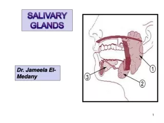
Anatomy • 3 Pairs – Major salivary glands • Parotid • Submandibular • Sublingual • Collection of salivary tissue within oral mucosa – Minor salivary glands www.nayyarENT.com
Development Of Parotid Gland • Ectoderm of oral cavity • Solid bulb from oropharyngeal epithelium • 6 weeks - parotid gland • Dichotomous branching of solid bulb, development of lumen, condensation of mesenchyme • Formation of primitive ducts www.nayyarENT.com
Contd… • Engulfment of facial nerve – 16th- 21st wk • Functional maturation after feeding is established www.nayyarENT.com
Parotid Gland • Lobulated, “inverted pyramid”, extent • Superficial, deep lobes • Parotid space • Borders - ant, post • Surfaces – superficial, superior, anteromedial, posteromedial www.nayyarENT.com
Capsule • Condensed deep cervical fascia, tough, inelastic surface component, thin deep layer • Stylomandibular ligament • Fibrous septa arise from capsule • Contents of fascia – superficial lymph nodes, greater auricular nerve www.nayyarENT.com
Structures Within The Gland • Facial nerve, division of gland • Retromandibular vein, anterior and posterior divisions • External carotid artery, terminal branches www.nayyarENT.com
Lymphoid Tissue In The Gland • Capsule – Periparotid Nodes • Mostly superficial to Facial Nerve • Part of MALT, secrete IgA • Salivary gland tissue may be present within the lymph nodes www.nayyarENT.com
Intraparotid Facial Nerve • Stylomastoid foramen • Methods of identification during surgery • TM Sulcus • PBD • Tragal pointer • Mastoid • Retrograde • Styloid process www.nayyarENT.com
Branching Patterns • Varied, Surgically important • Single trunk, divides into Zygomaticotemporal, Cervicomandibular • Temporal, upper / lower zygomatic, buccal • Buccal, cervical, mandibular www.nayyarENT.com
Contd… • Type1-5 ( Katz and Catalano, 1987) • Type 1 (25%) – No anastomotic links • Type 2 (14%) – Buccal fuses distally with Zygomatic • Type 3 (44%) – Major communication between Buccal & others • Type 4 (14%) – Anastomosis between major divisions • Type 5 (3%) – More than one Facial Nv trunk • Unpredictable preoperatively, to be precisely defined during surgery www.nayyarENT.com
Autonomic Nerve Supply Parasympathetic Inferior salivatory nucleus IX nerve Lesser Petrosal nerve Otic ganglion Auriculotemporal nerve PAROTID Sympathetic Superior cervical ganglion Plexus around ECA PAROTID www.nayyarENT.com
Parotid duct • Formed near the anterior border • Lies on superficial surface of Masseter • Opens in the mouth at parotid papilla • Accessory Parotid tissue www.nayyarENT.com
Submandibular Salivary Gland • Development • 6th IU wk • Ectoderm in floor of primitive oral cavity • Lateral to primitive tongue • Development of acini – 12th wk • Large superficial, small deep lobe • Located in Submandibular triangle • Well defined capsule www.nayyarENT.com
Superficial Lobe Inferior surface – Digastric, Deep fascia, Platysma, Skin Lateral surface – Submandibular fossa, Facial artery Surgical Anatomy • Medial surface – Mylohyoid, Hyoglossus, Lingual nerve, XII nv, Submandibular ganglion, Deep lingual vein www.nayyarENT.com
Deep Lobe • Extends for a variable distance between Mylohyoid & Hyoglossus • Relations • Superior – Lingual nerve • Inferior – XII Nv, Deep lingual vein, Submandibular duct www.nayyarENT.com
Wharton’s duct • 5 cm in length • Middle of deep part • Crosses Sublingual space • Proximally – b/w Mylohyoid & Hyoglossus • Distally – b/w Genioglossus & Sublingual gland • Opening – on sides of frenulum of tongue • Relation to Lingual nerve www.nayyarENT.com
Blood Supply & Lymphatic Drianage • Branches of Facial & Lingual arteries • Lymph nodes adjacent to the superficial part www.nayyarENT.com
Autonomic nerve supply • Parasympathetic Superior Salivary Nucleus NervusIntermedius Facial Nerve Chorda Tympani Lingual Nerve Submandibular Ganglion • Sympathetic Superior Cervical Ganglion Plexus around Facial Artery Submandibular Ganglion SUBMANDIBULAR GLAND www.nayyarENT.com
Surgery Of Submandibular Gland • Skin incision – 4 cm below Mandible • Ligation of Facial vessels above & below • Dissected away from Lingual Nerve • Lymph nodes in substance of gland www.nayyarENT.com
Sublingual Gland • Development • 8th wk • Epithelial buds present in paralingual sulcus • Almond shaped • Located in anterior part of floor of mouth www.nayyarENT.com
Relations Of Sublingual Gland • Sup – Oral floor mucosa • Inf – Mylohyoid • Post – Deep part Submandibular gland • Med – Lingual nerve, Submandibular duct, Genioglossus • Lat– Med surface of lower Mandible www.nayyarENT.com
Contd… • Ducts • Multiple • Drain into oral cavity directly or into Submandibular duct • Blood supply • Nerve supply www.nayyarENT.com
Physiology Of Salivary Glands www.nayyarENT.com
Function of Salivary Glands • Produce saliva – 1L / day (1ml/min/gm) • Contents • Mucin (glycoprotein) • Salivary amylase • Secretory Immunoglobulins • Other enzymes – DNase, RNase, lysozyme, lactoperoxidase, lingual lipase • Kallikerin • Inorganic compounds – Na+, K+, HCO3-, Ca2+ www.nayyarENT.com
Function Of Saliva • Lubrication and protection • Buffering and clearance • Maintenance of tooth integrity • Antibacterial activity • Taste and digestion www.nayyarENT.com
Structure of Salivary Gland • Parotid • Largest, serous (Compound Tubuloacinar Gland) • Submandibular and Sublingual • Mixed (Compound Tubuloacinar Glands) www.nayyarENT.com
Secretory End Pieces (Acini) • Serous Acini • Pyramid shaped, basal nucleus, apical secretory granules • Mucus Acini • Larger, columnar cells, basal nucleus • Mixed Acini • Mucus acini capped by serous cells forming Serous Demilunes www.nayyarENT.com
Duct System Acini Intercalated Ducts Striated Ducts Interlobular Excretory Ducts Stenson’s, Wharton’s duct www.nayyarENT.com
Control of Blood Flow And Metabolism • High rates • Rate of saliva production – 1ml/min/gm • Blood flow 10 times that of equal mass of skeletal muscle www.nayyarENT.com
Secretion Of Saliva • Active transport process under neuronal control • Composition • Hypotonic to plasma • Tonicity more when rates of production are high( at max rate - 70% to that of plasma) • K+,HCO3- higher than in plasma • pH – acidic during resting phase, basic during active phase(↑ HCO3- secretion) www.nayyarENT.com
Secretion Of Water And Electrolytes • Acini – Primary Fluid Secretion • Isotonic to plasma, electrolyte composition fairly constant, exocrine protein • Excretory ducts – extract Na+, Cl- and add K+, HCO3- to saliva • No addition in volume • More of Na+, Cl- removed than addition of K+, HCO3- responsible for hypotonicity www.nayyarENT.com
Mechanisms Of Primary Fluid Secretion • Osmotic process • Transepithelial salt gradients • Four ion transport systems - luminal and basolateral membranes generate the gradient • Three mechanisms proposed – operate concurrently www.nayyarENT.com
Mechanism 1 • Stimulation – rise in cytosolic Ca2+ • Opening of K+, Cl- channels – KCl outflow • Cl-conc in lumen ↑, Na+, H2O follow • Cl- entry sustained via Na+K+2Cl-cotransporter • 6 Cl-translocated to acinar lumen per ATP hydrolysed by Na+/K+ ATPase www.nayyarENT.com
Mechanism 2 • Cl-/HCO3-, Na+/H+ exchanger • KCl outflow • Cl- entry via Cl-/HCO3- exchanger • Acidification buffered by Na+/H+ exchanger • 3 Cl- translocated to lumen per ATP hydrolysed • Na+ & water follow into the lumen www.nayyarENT.com
Mechanism 3 • Involves acinar HCO3- secretion • 3 HCO3- secreted per ATP molecule • H+ extruded via Na+/H- exchanger • Na+, H2O follow into the lumen www.nayyarENT.com
Mechanism Of Macromolecule Secretion • Contained in zymogen granules present in serous acinar cells, ductal cells • Upon stimulation release contents in lumen by exocytosis • Conc and rate varies with level and type of stimulation www.nayyarENT.com
Mechanism Of Ductal Secretion • Inconstant, underlying mechanisms partially understood • Produce final hypotonic solution • Influence of tubular cells more when flow rate is slow www.nayyarENT.com
Neural Control Of Gland Function • Predominant control – PARASYMPATHETIC • Sympathetic stimulation shorter and less strong • Probable synergistic action www.nayyarENT.com
Parasympathetic Stimulation • Primary fluid secretion • Protein secretion • Vasodilatation • Increased metabolism and growth • Myoepithelial cell contraction LARGE VOLUME LOW PROTEIN OUTPUT www.nayyarENT.com
Sympathetic Stimulation • High protein secretion • Vasoconstriction – decreased blood flow • Myoepithelial cell contraction LOW VOLUME HIGH PROTEIN OUTPUT www.nayyarENT.com
Neurotransmitters & Receptors • Parasympathetic • Ach binds to M3 Receptors • Activation of G protein► Phospholipase C ►IP3 & DAG ► Intracellular Ca2+ release, Protein exocytosis www.nayyarENT.com
Contd… • Sympathetic • Noradrenaline binds to α1, β1 receptors • Activation of G protein ► Adenylate Cyclase activation ►↑cAMP dependant Protein Kinase ►protein exocytosis www.nayyarENT.com
Factors Affecting Salivary Flow • Unstimulated – Submandibular • Stimulated – Parotid 2/3rd • Acidic tastes – Max stimulation • Sweet tastes – Least stimulation www.nayyarENT.com
Contd… • Psychic factors • Circadian rhythm • Diurnal variation • Age • Drugs • Tricyclic antidepressants • Phenothiazines • Depression and anxiety states • Dehydration, hemorrhage, www.nayyarENT.com
Contd… • Salivary Gland diseases • Radiation sialadenitis • Autoimmune sialadenitis • HIV infection • Iron overload • Sarcoidosis • Amyloidosis • Cystic fibrosis www.nayyarENT.com
Factors Affecting Composition Of Saliva • Flow rate • Source of secretion • Type of stimulus • Diurnal variation • Diet • Drugs – flow dependant components • Hormones – mineralocorticoids, ovulation www.nayyarENT.com
Contd… • Disease states • Sialadenitis • Radiation damage • Sjorgen’s syndrome • Cystic fibrosis • HTN • DM • Alcoholic cirrhosis • Aldosteronism • Chronic pancreatitis www.nayyarENT.com
Salivary Assays In Diagnosis • Valid medium, painless, non-invasive • Hormone monitoring • Unconjugated steroids • Proportional to free unbound plasma levels • Useful in field studies • Estradiol, progesterone, testosterone www.nayyarENT.com