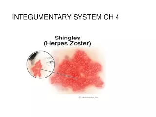
Chapter 4: Integumentary System
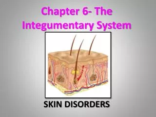
Chapter 4: Integumentary System. Integumentary System. What is it? The skin and its derivatives (sweat and oil glands, hair and nails) serve a number of functions, mostly protective. . Skin Structure. Figure 4.4. Skin Structure. Epidermis – outer layer Stratified squamous epithelium


Chapter 4: Integumentary System
E N D
Presentation Transcript
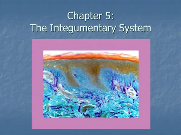
Integumentary System What is it? The skin and its derivatives (sweat and oil glands, hair and nails) serve a number of functions, mostly protective.
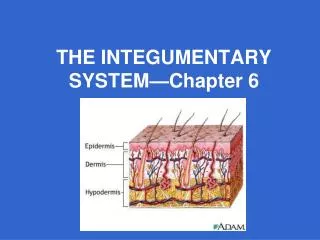
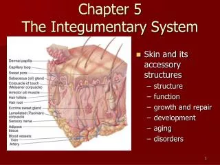
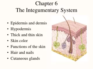
Skin Structure Figure 4.4
Skin Structure • Epidermis – outer layer • Stratified squamous epithelium • Often keratinized (hardened by keratin) • Dermis • Dense connective tissue Figure 4.3
Characteristics • 16% of your total body weight • First line of defense against microorganisms • Irradiated by sunlight • 1.5-2 M squared in area • There are 2 major components: 1) cutaneous membrane (skin) and 2) accessory structures (nails, hair and exocrine glands).
Keratinization • Or cornification, is the formation of protective , superficial layers of cells filled w/ keratin.
Functions of SKIN or Subcutaneous Membrane • Protection- of underlying tissues and organs from fluid loss, abrasion and impact and chemical attack and microbial infection. • Excretion- of salts, water and organic wastes and by integumentary glands. • Maintenance- of body temperature…via evaporation or insulation( Keratin) • Synthesis of Vitamin D3, a steroid, a hormone necessary for normal calcium absorption. • Detection of touch, pressure, pain…general senses that are relayed to the central nervous system.
SKIN: 2 TISSUES • 1. Epidermis- Contains either 4 or 5 layers depending on if THIN or THICK skin. • THIN: covers most of body ( 4 layers)(.08mm) • THICK: soles of feet, palms of hands (.5mm) (5 layers here. • Stratum: 1. germantivum(basale) 2. spinosum, 3.granulosum, 4. Lucidum (only in thick skin), and 5.corneum. • Example: Stratum germativum (basale)for 1st layer
Dermis:Components & Characteristics 2 COMPONENTS: • Papillary Layer- Consists of Areolar Tissue. Contains capillaries, lymphatics, and sensory neurons that supply the surface of the skin. Projections: Dermal Papillae. • Reticular Layer- deep to the PL, Contains blood vessels, deep pressure receptors called* Paciniancorpuscles and Macrophages. Also tissue : consists of interwoven layer of connective tissue; containing collagen and elastin fibers. Appendage structures extend into dermis.
DERMIS • Thicker than epidermis • Contains connective tissue; elastic fibers, collagen, nervous tissue, smooth muscle and blood;receptors and glands are found here. • A basement membrane separates the epidermis from dermis.
Papillary layer • loosely woven fibers • rich blood supply • irregular surface (fingerprints) Reticular layer • dense irregular CT • cleavage lines • less scarring • flexure lines—less sliding
Layers of Epidermis • Stratum basale (germinativum) • Stratum spinosum (prickly layer) • Stratum granulosum (granular) • Stratum lucidum (clear layer) • Stratum corneum (horny layer)
Stratum Basale • Single layer cells on basement membrane • Cell types in this layer • keratinocytes • undergo mitosis to replace epidermis • melanocytes • distribute melanin through cell processes • melanin picked up by keratinocytes • merkel cells are touch receptors • form Merkel disc
Melanin • Pigment (melanin) produced by melanocytes • Color is yellow to brown to black • Melanocytes are mostly in the stratum basale • Amount of melanin produced depends upon genetics and exposure to sunlight
These are found mostly in the stratum basale Everyone has the same number of melanocytes. The darker skin a person has depends on how much melanin a particular melanocyte produces and the size of the granules. This is determined by one’s genetics. UV rays can stimulate additional melanin production. Melanocytes
Melanocyte Stimulation • Melanocytes deeper in the epidermis are stimulated to produce new melanin granules. These new granules are transferred to keratinocytes in the upper cell layers of the skin. These granules become positioned in the outer portions of the cells above the cell nuclei, thus providing additional protection.
If Melanocytes on Bottom, How Does Skin Get Tan on Top? • Melanocytes have extensions that transfer the granules to surface cells by a process known as Cyocrine Secretion.
Stratum Spinosum • Contain special white blood cells from the lymphatic system. Help protect body from pathogens.
Statum Grandulosum • Produces lipid-filled vesicles thatrelease a glycolipid by exocytosisto waterproof the skin
Statum Lucidum • Thin translucent zone seen only in thick skin ( palms and soles of feet) • Cells have no nucleus or organelles
Stratum Corneum • The most superficial layer of epidermis • 20- 30 layers thick • Cornified, ABSOLUTELY dead • ABSOLUTELY flattened • These take up 75% of the epidermis. • They exfoliate continuously.
Appendage Structures Cutaneous Glands- the EXOCRINE glands, including sweat and sebaceous glands. • Both of these glands are formed in epidermis and pushed down during development and remain almost entirely in dermis. • Sebaceous- OIL glands. Usually empty into hair follicle, sometimes directly skin. ---SEBUM=oil and skin cells, germicide.
Named after discovered Theses are sensory receptors that detect Pacinian Receptors
Sebaceous (Oil) Glands Secrete oil or sebum Everywhere except palms, soles Usually secretes into hair follicle Lubricates hair and skin • softens dead cells--pliability • slows water loss • bactericidal Stimulated by hormones (androgens)
Sebaceous glands associated with hair follicle Sweat gland
sss Sebaceous glands associated with hair follicle Eccrine glands , that is SUDORIFEROUS or SWEAT GLAND empties onto Skin
Apendages (cont.) • Sweat Glands- Eccrine and Apocrine • Eccrine- more numerous, ubiquitious, used to regulate body temperature.Empty onto skin. • Apocrine- found auxillary, and pubic areas only. Develop during puberty under influence of testosterone. Contain protein and fat. Do not regulate body temp. Activated by stress and sexual foreplay. Empty onto hair follicle.. usually.
What Did You Say??? • Ceruminous glands- This are modified (eccrine) sweat glands in the passageway of the external ear. Their secretions combine w/ sebaceous glands forming CERUMEN….or earwax. Together w/ tiny hairs traps foreign particles.
Subcutaneous Layer • Located under the dermis • Mostly adipose • Functions • energy reservoir • thermal insulation • Hypodermic injections • highly vascular
Skin Cancer • Skin cancer is the most common type of cancer in humans. Cause is unknown, but most important risk factor is over exposure to UV light. Frequent irritation to skin by infections, chemicals or physical trauma seem to be a predisposing. Also Risk factors: living at high altitudes, fair skinned people, having a lot of moles, a family history of the disease and if you had one or more severe sunburns as a child
Skin Cancer: RFs • over exposure to UV light. • Frequent irritation to skin by infections, chemicals or physical trauma • living at high altitudes, • fair skinned people, • having a lot of moles • family history of the disease • one or more severe sunburns as a child
Skin Cancer: 3 Types • Basal – arise from the layer stratum basale. Least malignant, most common. Cannot form keratin ( soft). Looks like a mound. No boundary between epidemis and dermis. • Squamous – arise from statum spinosum. Characterized by pearly beaded edge. These will metastasize into the lymph nodules if left untreated and can be lethal. • Melanoma- rarest but most deadly. Develops from pigmented moles. 5% survival rate.
ABCD Test For Melanoma A – Asymmetrical; the two sides of the pigmented spot do not match. B- BORDERS-irregular boarders; not smooth with indentations apparent C- COLOR- pigment contains areas of different colors D- DIAMETER- larger than ¼ inch
Burns • Hot water, sunlight, radiation, electric shock or acids and bases • Death from fluid loss and infection • Degrees of burns • 1st-degree = only the epidermis (red, painful and edema) • 2nd-degree = epidermis and part of dermis (blistered) • epidermis regenerates from hair follicles and sweat glands • 3rd-degree = epidermis, dermis and more is destroyed • often requires grafts or fibrosis and disfigurement may occur • Treatment – IV nutrition and fluid replacement, debridement and infection control