Download
1 / 10
110 likes | 215 Vues
Explore the intricate processes of heart conduction and the cardiac cycle. Learn how the heart's rhythmic beat is controlled, the role of the autonomic nervous system, and the phases of the cardiac cycle. Understand the importance of the SA node, atrial and ventricular contractions, and the circulation of blood through the heart.

E N D
Cardiac Circulation • Myocardium nourished by coronary artery • Coronary veins drain into coronary sinus (enlarged vessel on backside of heart) • Coronary sinus empties into right atrium
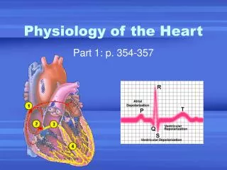
Heart Conduction • Cardiac muscle does not have to be controlled by nervous system • Muscle cells in different areas of heart have different rhythms
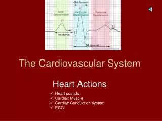
Heart Rate influences • Autonomic nervous system increases & decreases heart rate • Intrinsic conduction system (nodal system) causes heart muscle depolarization (electric impulse that causes heart beat)
Heart Beat • SA node (also known as pacemaker) • Located in right atrium • Starts heart beat • Impulse travels through atrium to the AV node • Atrium contract
Impulse through AV bundle through bundle branches to Purkinje fibers in apex • Ventricle contracts starting at apex and moving toward the atria. • Blood forced upward out of the aorta
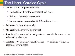
Cardiac Cycle = complete heart beat • Atria contract simultaneously • Atria relax • Ventricles contract • Systole = contraction of ventricle • Diastole = relaxation of ventricle
Mid to late diastole • Ventricle completely relaxed • Pressure low in heart • Blood flowing passively into and through atria into ventricles • Semilunar valves closed, AV valves open • Then atria contract, forcing blood into ventricles
2. Ventricular systole • Ventricles contract (atria relax-filling with blood) • Pressure increases, closing AV valves (lub sound) • Higher pressure in ventricles, than in arteries so forces semilunar valves open and blood is forced into arteries
3. Early diastole • Ventricles relax • Semilunar valves close (preventing back flow) • Dup sound • Intraventicular pressure drops, until low enough for AV valves to be forced open