Download
1 / 31
350 likes | 679 Vues
Organization of DNA. Viral DNA (and in some, RNA) Some circular, some linear Some double stranded, some single stranded Very small amount, packed very tightly Small size is an advantage Viruses use host cell enzymes, need few genes Bacterial DNA Usually single copy of double stranded

E N D
Organization of DNA • Viral DNA (and in some, RNA) • Some circular, some linear • Some double stranded, some single stranded • Very small amount, packed very tightly • Small size is an advantage • Viruses use host cell enzymes, need few genes • Bacterial DNA • Usually single copy of double stranded • Usually circular • Eukaryotic DNA: linear, in several pieces
DNA packaging For example, the chromosome of E. coli is 1.2 mm long, but must fit into a bacterium that is only 0.001 mm long! http://www.expatica.com/xpat/xpatsite/www/upload_pix/surprised-face.jpg
Protein packaging of DNA • Four proteins in E. coli • Make up 10% of total protein of cell • HU for wrapping; FIS and IHF for bending; HNS for compaction. • Same function as histone proteins in eukaryotes. • Positively charged proteins bind to negatively charged DNA. • End result: nucleoid, a region in the cytoplasm rich in DNA and protein; comparable to a nucleus but without a membrane.
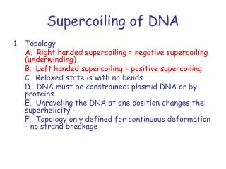
DNA of E. coli is supercoiled • In addition to being packaged with proteins, the DNA of E. coli is supercoiled. • Supercoiling. DNA could be “relaxed” or supercoiled. In Eubacteria, DNA is “underwound” (negatively supercoiled); • Supercoiling carried out by topoisomerases. • Example: gyrase, that relieves stress during DNA replication. • Two types, depending on whether 1 or two DNA strands are cut (and repaired) in the process.
Supercoiling Top left: relaxed DNA Bottom left: supercoiled. Bottom: schematic of underwinding DNA.
Packaging of E. coli DNA Note arrows: one shows where the DNA has been “nicked”, relaxing the supercoiling. The other points to a supercoiled region. That supercoiling can be relaxed in ONE PLACE means that the DNA is constrained in places.
The enslaved bacteria • Mitochondria and chloroplasts thought to have originated as prokaryotic endosymbionts in early eukaryotes • Carry out respiratory functions in membrane • DNA is circular, ds DNA like in prokaryotes • Self replicating • Have their own ribosomes, similar to bacterial • Organelle DNA discovered from mutations • Some traits not determined by nuclear genes • Inheritance via mother; ovum has all the cytoplasm
Integration of organelles is thorough • Mitochondria: • Replication requires nuclear genes • Polymerases, initiation factors, respiratory proteins are multi-subunit proteins • Several of the subunits for each are nuclear, others are mitochondrial • Chloroplasts • Multi-subunit enzymes jointly encoded • Genes for RuBP carboxylase divided between nucleus and chloroplast
Polytene chromosomes Occur in the salivary glands of various flies during development. Condensed areas of DNA line up, produce darkly staining bands. Useful for mapping genes: banding patterns are unique, and in situ hybridization can be used to localize genes on DNA
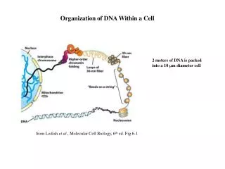
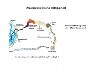
DNA packaging in eukaryotes • Largest human chromosome is made of DNA which is 82 mm long (over 3 inches) • During metaphase, DNA is further compacted to about 10 µm long. • Equivalent to winding 25 miles of spaghetti into a 16 foot canoe. • DNA has to be well packaged to fit into the cell, to be compacted even more during mitosis • still has to be accessible during interphase for use! • Chromatin: grainy appearing mixture of DNA and proteins in the nucelus
Nucleosomes: unit of packaging of eukaryotic DNA DNA wrapped around histone proteins: TWO each of the proteins H2A, H2B, H3, and H4. Additionally, H1 on outside helps hold DNA to structure.
About histones and arrangement • Histones • positively charged, to attach well to DNA • conserved, very little difference among organisms • How arrangement was determined • DNA collected, treated briefly with nuclease to see how much DNA is protected by proteins • Remove proteins, separate DNA pieces by size on gel • 200 bp pieces of DNA produced • treat more with nuclease, repeat analysis • get 145 bp DNA pieces
Structure deduced • the 145 bp of DNA are wrapped around the histone octet which is the core particle. • 200 bp includes region covered by H1 which covers DNA as it enters, exits nucleosome. • the rest of the DNA is linker DNA between.
Nucleosomes are wound up Fig. 8.9 shows how “beads on a string” are further wound up to produce a solenoid, the structure of chromatin. During mitosis, this solenoid itself coils further to make chromatids.
Organization of DNA • Does DNA packaging create problems? • DNA wrapped tightly around histones • DNA must be accessible for replication, transcription • Modification of histones changes packing with DNA • Acetylation: acetyl groups added to histones. • Phosphorylation: phosphate groups added by kinases • These groups decrease positive net positive charges, allow DNA freedom.
Differences in DNA • Heterochromatin vs. Euchromatin • Heterochromatin is DNA which tends to be highly compacted and dark staining. • Euchromatin is not so compacted or dark. • The number of genes in heterochromatin is generally small relative to euchromatin. • Heterochromatin lacks genes or they are inactive • Much heterochromatin is found in certain structural parts of the chromosomes: centromeres and telomeres. Also, much of Y chromosome. • Move euchromatin to an area next to heterochromatin and it becomes heterochromatin: position effect.
Chromosome structure Arm http://www.med.uiuc.edu/m1/genetics/images/webun1/Chromosome.gif medic.med.uth.tmc.edu/.../ cellbio/hist-01.htm
Composition of DNA:% G+C There is always equal #s of A and T, and G and C, but the percentage of G+C pairs and A+T pairs can be different among different organisms.
Measuring % G+C hyperchromic shift As DNA “melts”, becomes SS, absorbs more UV at 260 nm. Because G-C pairs have 3 H-bonds instead of two, DNA with more G+C is more stable, melts at higher temperature (blue).
Satellite DNA • In prokaryotes, the %G+C base pairs is pretty much averaged out over the entire DNA; not so with eukaryotes. • Density gradient ultracentrifugation can also be used to determine %G+C. • G+C pairs are denser than A+T, migrate to a lower location (greater density) in the gradient. • Fragmented eukaryotic DNA showed something odd…
Satellite DNA When the DNA was analyzed, a portion has a lower %G+C than the rest of the DNA, producing a “satellite band”. How could a portion of DNA have a different composition than the rest?
Repeated sequences • If a section of DNA with a %G+C composition different from the rest of the DNA is repeated many times, DNA fragments from these regions of DNA would behave differently during the centrifugation.
Study of the Composition of DNA using DNA renaturation kinetics • Break DNA into random fragments. • Denature with heat (melt). • Cool, allow strands to find their complements and go from ss to ds again (anneal). • Follow entire process using UV light absorption at 260 nm • as DNA goes from ss to ds, Abs decreases.
Renaturation kinetics • Kinetics: study of the rate of change. • Major Point #1: the more copies of the complementary strands there are, the less time they will take to find each other • the more DNA, the faster the process. In this fig., 2 different amounts of DNA from the SAME organism.
Renaturation kinetics-2 • Major Point #2: • Given equal amounts • (same mass) of DNA, • the bigger the total genome • of the organism, the slower • the renaturation. • If the genome is bigger, and the amounts of DNA used in the experiment are the same, the organism with the bigger genome will have fewer copies of the complementary fragments, so annealing will take longer (see point #1).
Understanding genome size Imagine you have 20 playing cards. In one instance, you have these 5 cards, another 5 cards exactly the same, and 2 more sets of the Ace thru 10 but of diamonds. In the second instance, you have ace thru 5 of hearts and also of diamonds. In which case will you match up pairs of hearts and diamonds most quickly? The smaller deck gets matched up quicker. http://www.skydiveelsinore.com/calendar/images/playing-cards-spread.jpg
Cot curves: Studying renaturation of DNA The amount of DNA affects the rate at which DNA fragments renature. To avoid the problem of comparing samples with different amounts of DNA, the change in ss DNA is graphed vs.the initial DNA concentration (Co) x the time (t): Cot Y-axis is the fraction or percent of the DNA that is ss (experiment starts by denaturing the DNA). X-axis is Cot which is a Log scale. www.cas.muohio.edu/.../gene2000/ lect7/fig9p8c.jpg
Cot curves and satellite DNA Categories variable among different organisms. Highly repetitive DNA, many complements, find each other quickly. Single copy (unique sequence) much slower. http://www.ndsu.nodak.edu/instruct/mcclean/plsc431/eukarychrom/cot2.gif
Types of DNA • Unique, single copy: typically 30-75% of DNA in most eukaryotes. • Highly repetitive DNA: 5-45 % of DNA depending on species. In humans: • ALU family: contains Alu I site. 300 bp long, appears 500,000 times, dispersed. 5% of DNA. • SINEs = short intersperesed elements • transposable • Alpha satellite DNA: tandem repeats of 170 bp occur 5,000-15,000 times; make up part of centromere. 6% • L1 family (in humans), example of LINEs • Long interspersed elements • transposable
Middle or moderately repetitive DNA • Moderately repetitive DNA: • Tandem or interspersed repeats • VNTRs, good for DNA fingerprinting • Variable number tandem repeats • 15 – 100 bp long, between or within genes • Dinucleotide repeats (CA)N, also good for forensic work • in maize and yeasts: transposons in large numbers. • genes for rRNA, tRNA, ribosomal proteins, histones
All your DNA codes for proteins?Sorry, not close • Only 4% codes for proteins, in 30,000 genes • 96% of DNA includes • Introns, “junk” DNA within and around genes. • Genes coding for rRNA and tRNA • Junk DNA called repetitive sequences • Pseudogenes; have sequences that look like genes but are never expressed, don’t work. • We are related to everything else • Our genes look like those from chimpanzees, bacteria.