Download
1 / 53
540 likes | 747 Vues
Organization and Synthesis of DNA. Andy Howard Introductory Biochemistry 15 October 2009. Restriction Enzymes (concluded) Review of A,B,Z DNA Intercalation Denaturation and renaturation of DNA DNA density. DNA tertiary structure Review of supercoiling Gyrases Nucleosomes Higher levels

E N D
Organization and Synthesis of DNA Andy HowardIntroductory Biochemistry15 October 2009 Biochemistry:Nucleic Acids III
Restriction Enzymes (concluded) Review of A,B,Z DNA Intercalation Denaturation and renaturation of DNA DNA density DNA tertiary structure Review of supercoiling Gyrases Nucleosomes Higher levels Bacterial organization What we’ll discuss Biochemistry:Nucleic Acids III
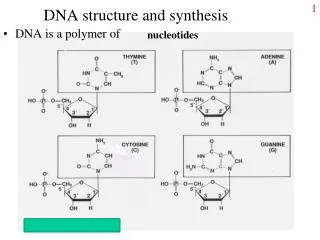
The biology problem • How does the bacterium mark its own DNA so that it does replicate its own DNA but not the foreign DNA? • Answer: by methylating specific bases in its DNA prior to replication • Unmethylated DNA from foreign source gets cleaved by restriction endonuclease • Only the methylated DNA survives to be replicated • Most methylations are of A & G,but sometimes C gets it too Biochemistry:Nucleic Acids III
How this works • When an unmethylated specific sequence appears in the DNA, the enzyme cleaves it • When the corresponding methylated sequence appears, it doesn’t get cleaved and remains available for replication • The restriction endonucleases only bind to palindromic sequences Biochemistry:Nucleic Acids III
Use of restriction enzymes • Nature made these to protect bacteria; we use them to cleave DNA in analyzable ways • Similar to proteolytic digestion of proteins • Having a variety of nucleases means we can get fragments in multiple ways • We can amplify our DNA first • Can also be used in synthesis of inserts that we can incorporate into plasmids that enable us to make appropriate DNA molecules in bacteria Biochemistry:Nucleic Acids III
Summaries of A, B, Z DNA Biochemistry:Nucleic Acids III
DNA is dynamic • Don’t think of these diagrams as static • The H-bonds stretch and the torsions allow some rotations, so the ropes can form roughly spherical shapes when not constrained by histones • Shape is sequence-dependent, which influences protein-DNA interactions Biochemistry:Nucleic Acids III
Intercalating agents • Generally: aromatic compounds that can form -stack interactions with bases • Bases must be forced apart to fit them in • Results in an almost ladderlike structure for the sugar-phosphate backbone locally • Conclusion: it must be easy to do local unwinding to get those in! Biochemistry:Nucleic Acids III
Instances of inter-calators Biochemistry:Nucleic Acids III
Denaturing and Renaturing DNA See Figure 11.17 • When DNA is heated to 80+ degrees Celsius, its UV absorbance increases by 30-40% • This hyperchromic shift reflects the unwinding of the DNA double helix • Stacked base pairs in native DNA absorb less light • When T is lowered, the absorbance drops, reflecting the re-establishment of stacking Biochemistry:Nucleic Acids III
Heat denaturation • Figure 11.14Heat denaturation of DNA from various sources, so-called melting curves. The midpoint of the melting curve is defined as the melting temperature, Tm.(From Marmur, J., 1959. Nature183:1427–1429.) Biochemistry:Nucleic Acids III
GC content vs. melting temp • High salt and no chelators raises the melting temperature Biochemistry:Nucleic Acids III
How else can we melt DNA? • High pH deprotonates the bases so the H-bonds disappear • Low pH hyper-protonates the bases so the H-bonds disappear • Alkalai is better: it doesn’t break the glycosidic linkages • Urea, formamide make better H-bonds than the DNA itself so they denature DNA Biochemistry:Nucleic Acids III
What happens if we separate the strands? • We can renature the DNA into a double helix • Requires re-association of 2 strands: reannealing • The realignment can go wrong • Association is 2nd-order, zippering is first order and therefore faster Biochemistry:Nucleic Acids III
Steps in denaturation and renaturation Biochemistry:Nucleic Acids III
Rate depends on complexity • The more complex DNA is, the longer it takes for nucleation of renaturation to occur • “Complex” can mean “large”, but complexity is influenced by sequence randomness: poly(AT) is faster than a random sequence Biochemistry:Nucleic Acids III
Second-order kinetics • Rate of association: -dc/dt = k2c2 • Boundary condition is fully denatured concentration c0 at time t=0: • c / c0 = (1+k2c0t)-1 • Half time is t1/2 = (k2c0)-1 • Routine depiction: plot c0t vs. fraction reassociated (c /c0) and find the halfway point. Biochemistry:Nucleic Acids III
Typical c0t curves Biochemistry:Nucleic Acids III
Hybrid duplexes • We can associate DNA from 2 species • Closer relatives hybridize better • Can be probed one gene at a time • DNA-RNA hybrids can be used to fish out appropriate RNA molecules Biochemistry:Nucleic Acids III
GC-rich DNA is denser • DNA is denser than RNA or protein, period, because it can coil up so compactly • Therefore density-gradient centrifugation separates DNA from other cellular macromolecules • GC-rich DNA is 3% denser than AT-rich • Can be used as a quick measure of GC content Biochemistry:Nucleic Acids III
Density as function of GC content Biochemistry:Nucleic Acids III
Tertiary Structure of DNA • In duplex DNA, ten bp per turn of helix • Circular DNA sometimes has more or less than 10 bp per turn - a supercoiled state • Enzymes called topoisomerases or gyrases can introduce or remove supercoils • Cruciforms occur in palindromic regions of DNA • Negative supercoiling may promote cruciforms Biochemistry:Nucleic Acids III
DNA is wound • Standard is one winding per helical turn, i.e. 1 winding per 10 bp • Fewer coils or more coils can happen: • This introduces stresses that favors unwinding • Both underwound and overwound DNA compact the DNA so it sediments faster than relaxed DNA Biochemistry:Nucleic Acids III
Linking, twists, and writhe • T=Twist=number of helical turns • W=Writhe=number of supercoils • L=T+W = Linking number is constant unless you break covalent bonds Biochemistry:Nucleic Acids III
Examples with a tube Biochemistry:Nucleic Acids III
How this works with real DNA Biochemistry:Nucleic Acids III
How gyrases work • Enzyme cuts the DNA and lets the DNA pass through itself • Then the enzyme religates the DNA • Can introduce new supercoils or take away old ones Biochemistry:Nucleic Acids III
Typical gyrase action • Takes W=0 circular DNA and supercoils it to W=-4 • This then relaxes a little by disrupting some base-pairs to make ssDNA bubbles Biochemistry:Nucleic Acids III
Superhelix density • Compare L for real DNA to what it would be if it were relaxed (W=0): • That’s L = L - L0 • Sometimes we want = superhelix density= specific linking difference = L / L0 • Natural circular DNA always has < 0 Biochemistry:Nucleic Acids III
< 0 and spools • The strain in < 0 DNA can be alleviated by wrapping the DNA around protein spool • That’s part of what stabilizes nucleosomes Biochemistry:Nucleic Acids III
Cruciform DNA • Cross-shaped structures arise from palindromic structures, including interrupted palindromes like this example • These are less stable than regular duplexes but they are common, and they do create recognition sites for DNA-binding proteins, including restriction enzymes Biochemistry:Nucleic Acids III
Cruciform DNA example Biochemistry:Nucleic Acids III
Eukaryotic chromosome structure • Human DNA’s total length is ~2 meters! • This must be packaged into a nucleus that is about 5 micrometers in diameter • This represents a compression of more than 100,000! • It is made possible by wrapping the DNA around protein spools called nucleosomes and then packing these in helical filaments Biochemistry:Nucleic Acids III
Chromatin • Discovered long before we understood molecular biology • Seen to be banded objects in nuclei of stained eukaryotic cells • In resting cell it exists as long slender threads, 30 nm diameter From answers.com Biochemistry:Nucleic Acids III
Squishing the DNA • If the double helix were fully extended, the largest human chromosome (2.4*108bp) would be 2.4*108 *0.33nm ~ 0.8*108nm=80 mm; • much bigger than the cell! • So we have to coil it up a lot to make it fit. • Longest chromosome is 10µm long • So the packing ratio is 80mm/10µm = 8000 Biochemistry:Nucleic Acids III
Chromosome structure: levels • Each of the first 4 levels compacts DNA by a factor of 6-20; those multiply up to > 104 Biochemistry:Nucleic Acids III
Nucleosome Structure • Chromatin, the nucleoprotein complex, consists of histones and nonhistone chromosomal proteins • Histone octamer structure has been solved • without DNA: Moudrianakis, 1991 • with DNA by Richmond • Nonhistone proteins are regulators of gene expression Biochemistry:Nucleic Acids III
Histone types • H2a, H2b, H3, H4 make up core particle: two copies of each, so: octamer • All histones are KR-rich, small proteins • H1 associates with the regions between the nucleosomes Biochemistry:Nucleic Acids III
Histones: table 11.2, plus… Biochemistry:Nucleic Acids III
Unfolded chromatin • Treat chromatin with low ionic strength; that disrupts higher level interactions so the individual nucleosomes are strung out relative to one another like beads on a string Image courtesy U. Maine Biochemistry:Nucleic Acids III
Nucleosome core particle Biochemistry:Nucleic Acids III
Half the core particle • Note that DNA isn’t really circular: it’s a series of straight sections followed by bends Biochemistry:Nucleic Acids III
Histones, continued • Individual nucleosomes attach via histone H1 to seal the ends of the turns on the core and organize 40-60bp of DNA linking consecutive nucleosomes • N-terminal tails of H3 & H4 are accessible • K, S get post-translational modifications, particularly K-acetylation Biochemistry:Nucleic Acids III
Histone deactivation • Histones interact with DNA via +charges on lys and arg residues. • If we neutralize those charges by acetylation, the histones don’t bind as tightly to the DNA • Carefully-timed enzymatic control of histone acetylation is a crucial element in DNA organization Biochemistry:Nucleic Acids III
CoASH Histone acetylation Histone H1PDB 1GHC8.3 kDa monomerChicken • Active histone + Acetyl CoA inactive (acetylated) histone + CoASH • Without the positive charges, the affinity for DNA goes down Histone acetyltransferasePDB 1QSO66 kDatetrameryeast Biochemistry:Nucleic Acids III
Histone deacetylation • Type III deacetylases usea non-trivial reaction:Prot-lys-NAc + NAD+ Prot-lys-NH3+ + nicotinamide +2’-O-acetyl-ADP-ribose • Part of the NAD salvage pathway Histone/protein deacetylase +histone H4 active peptidePDB 1SZD; 34 kDa “heterodimer”yeast Biochemistry:Nucleic Acids III
Nucleosome structure • Core octamer is two molecules each of H2A, H2B, H3, H4 • Typically wraps around~200bp of DNA • DNA betweennucleosomes is ~54 bp long • H1 binds to linker and to core particle; but in beads-on-a-string structure, it’s often absent Biochemistry:Nucleic Acids III
How much does this coil up? • 200 bp extended would be about 50nm • The width of the core-particle disk is 5nm • So this is a tenfold reduction • Nucleosomal organization corresponds to negative supercoiling • … so DNA ends up supercoiled when we take away the histones Biochemistry:Nucleic Acids III
Courtesy answers.com Next level of organization • H1 interacts with DNA along linker region • Individual histones spiral along to form 30 nm fiber • See fig.19.25 Courtesy Johns Hopkins Univ Biochemistry:Nucleic Acids III
Even higher… • The 30nm fibers are attached to an RNA-protein scaffold that holds the 30nm fibers in large loops • Typical chromosome has ~200 loops • Loops are attached to scaffold at their base • Ends can rotate so it can be supercoiled Biochemistry:Nucleic Acids III