PANCREATIC HORMONES
1.56k likes | 3.83k Vues
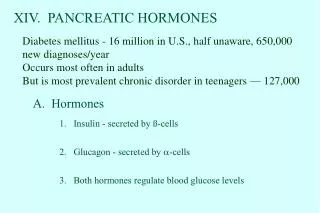
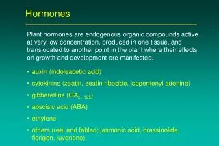
PANCREATIC HORMONES. OBJECTIVES. To know the Functional Anatomy of Pancreas To be able to differentiate b/w exocrine & endocrine functions of Pancreas To be able to enlist the hormones produced by the pancreas & the pancreatic cells that produce them

PANCREATIC HORMONES
E N D
Presentation Transcript
OBJECTIVES • To know the Functional Anatomy of Pancreas • To be able to differentiate b/w exocrine & endocrine functions of Pancreas • To be able to enlist the hormones produced by the pancreas & the pancreatic cells that produce them • To be able to define different terms used for carbohydrate metabolism • To know the structure of Insulin. • To know the synthesis of Insulin • To know the actions of Insulin • To be able to tell the MOA of Insulin
PANCREAS Pancreas is an elongated organ with one end broad (shaped like a hook) & the other end narrowing to a tail. This organ lies sideways, the hook on the right side and turned downwards. It lies behind & below the stomach fitted in the C-shaped concavity of the loop of duodenum (small intestine)
FUNCTIONAL ANATOMY OF PANCREAS • Pancreas is a mixed gland that performs both exocrine & endocrine functions • WHAT IS THE DIFFERENCE B/W EXOCRINE & ENDOCRINE GLANDS?
FUNCTIONAL ANATOMY OF PANCREAS EXOCRINE TISSUES Larger part Grape-like clusters of secretory cells forming sacs called ACINI Which empty into the pancreatic ducts that eventually empty into the duodenum The secretion is called the Pancreatic juice!— What does the pancreatic juice contain?
FUNCTIONAL ANATOMY OF PANCREAS ENDOCRINE TISSUES Smaller part consists of isolated islands of endocrine tissues called as ISLETS OF LANGERHANS dispersed throughout. It forms only 1% of the total pancreatic mass. 1-2 million in number 0.3 mm in diameter These secrete HORMONES!
HORMONES OF PANCREAS Enlist the hormones of Pancreas: Insulin (also Proinsulin & C-peptide) Glucagon Amylin Somatostatin Pancreatic peptide
STRUCTURE of INSULIN It is a Peptide hormone. It consists of 2 amino acid (51AA) chains that are joined together by disulphide linkages. If the 2 AA chains are split apart then the functional activity of insulin is lost. It has a molecular weight of 5808.
Synthesis of Insulin • Synthesis of Insulin occurs in the rough endoplasmic reticulum of β- cells in Islets of Langerhans. • It is initially produced as a Preprohormone called Preproinsulin (mw: 11,500) which is cleaved in the ER to yield Proinsulin (mw: 9,000). • Proinsulin is further cleaved in Golgi apparatus to yield Insulin and its peptide fragment which is also called the C-peptide (connecting peptide). • C-peptide is a connecting peptide that connects α and β chains. • These both are then packaged in secretory vesicles & released when the stimulus arrives.
Synthesis of Insulin Preproinsulin (ER) (11,500) ↓ Proinsulin (Golgi Apparatus) (9,000) ↓ cleavage Insulin + C-peptide (stored in secretory vesicles) ↓ Secreted into blood ↓ After use degraded by the enzyme called as Insulinase in the liver & to a lesser extent in the kidneys & muscles
Insulin Metabolism • Plasma half-life: 6 minutes • Cleared from the circulation in 10-15 minutes • However, the C-peptide takes about 30 Minutes to be degraded. Advantage?
Insulin Secretion Insulin release is not continuous even after a meal but oscillates with a period of 3-6 minutes: spurts of insulin release more than 800 pmol/l to less than 100 pmol/l. This oscillation is important to consider when administering insulin-stimulating medication as oscillation is the target & not a constant high concentration.
INSULIN RECEPTOR It is a tetramer formed by 4 glycoprotein subunits: 2 alpha subunits (present outside the cell membrane) 2 beta subunits (penetrate through the memb. Into the cell cytoplasm) The alpha and beta subunits are linked by disulfide bonds. It is an enzyme-linked receptor. Contains multiple enzyme groups called as Insulin-receptor substrates (IRS). Different types of IRS are expressed in different tissues (IRS1-3).
Insulin receptor activation Insulin attaches to alpha subunit of receptor ↓ The beta subunit of the receptor becomes autophosphorylated ↓ Tyrosine kinase is activated ↓ IRS is Phosphorylated. ↓ Action exerted e.g. glucose uptake by the cells is enhanced.
Mechanism of Insulin secretion Glucose binds to GLUT-2 in the beta cells of Islets of Langerhans and enters the cell. ↓ Glucokinase phophorylates Glucose to G-6-phosphate ↓ G-6-phosphate is oxidized to form ATP ↓ ATP inhibits the ATP-sensitive K channels of the cell ↓ Closure of the K channel leads to depolarization of the cell membrane ↓ Voltage-gated Ca channels are opened ↓ Ca influx takes place ↓ Stimulates fusion of insulin-containing vesicles with cell membrane ↓ Exocytosis of Insulin to the ECF
WHAT IS THE OBJECTIVE OF INSULIN? ↓ It is to maintain blood glucose homeostasis!
Normally, circulating glucose concentration is determined by:
POINT TO REMEMBER! • INSULIN IS THE ONLY HORMONE CAPABLE OF LOWERING THE BLOOD GLUCOSE LEVEL.
INSULIN • Insulin is an ANABOLIC hormone. • Insulin is the hormone of the FED/ ABSORPTIVE state. • Insulin has important effects on: - CHO - Fats - Proteins • It LOWERS blood glucose levels of: - Glucose - fatty acids - amino acids • It is a hormone associated with ENERGY ABUNDANCE.
Lack of effect of Insulin on Glucose uptake by BRAIN • Brain uses ONLY Glucose as its energy source, therefore, it is important that blood glucose levels be maintained above a critical level. • Brain is PEREMEABLE to Glucose & can use it even without the intermediation of Insulin. • When blood glucose levels fall too low ( 20-50mg/ 100ml), symptoms of hypoglycemic shock develop. • Hypoglycemic shock is characterized by progressive nervous irritability that leads to fainting, seizures & even coma
Insulin increases glucose transport into most, but not all, insulin- sensitive cells. Role of GLUT: The transport of glucose b/w blood and different tissue cells is accomplished by the proteins called GLUCOSE TRANSPORTERS (GLUT). • Fourteen different Glucose transporters have been characterized, GLUT 1-14 in the order of discovery. • The GLUT all accomplish passive diffusion of Glucose across the cell membrane. Once inside the cell, the Glucose is immediately phosphorylated to Glucose-6-Phosphate which cannot leave the cell through the bi-directional GLUT protein and is “trapped” inside the cell. • Each GLUT has been evolved for a different task & a different tissue. • GLUT 1,2,3 & 5 are NOT affected by insulin: - GLUT-1: transports glucose across blood brain barrier - GLUT-2: from kidney & intestinal cells into the blood stream. Glucose enters the kidney & intestinal cells by SGLT (Na & Glucotransporters) - GLUT-3: neurons - GLUT-4: in all major cells using Glucose (as muscle and adipose tissues) Therefore, in all these tissues the glucose entry is insulin independent.
GLUT-4 • Only GLUT-4 is insulin-dependant & occurs in the muscles & adipocytes. • These cells maintain a pool of GLUT-4 molecules in vesicles in their cell cytoplasm.
GLUT-4 Insulin attaches to the receptor ↓ Insulin receptor is activated ↓ Vesicles containing GLUT-4 move rapidly to the cell membrane ↓ Vesicles fuse with the cell membrane, inserting the transporter in the membrane ↓ When insulin action ceases, the transporter-containing patches of membrane are endocytosed ↓ Vesicles are reformed ready for the next action
ROLE OF GLUT-4 In the absence of Insulin, Glucose cannot enter the cell Insulin signals the cell to insert GLUT 4 transporters into the membrane, allowing glucose to enter the cell.
Glucose entry into the LIVER • Glucose does not depend on GLUT-4 for entry into the LIVER.
Insulin stimulates Glucose uptake by the cells (mostly thru GLUT-4). • Insulin stimulates Glycogenesis in both the Liver & the Skeletal muscles. • It inhibits Glycogenolysis. • It inhibits Gluconeogenesis. • It promotes liver uptake, use & storage of Glucose
Action on CHO met. In MUSCLES Throughout the day, the muscles use Fatty Acids as an energy source EXCEPT: - During moderate & heavy exercise when they uptake Glucose through Insulin-independent pathway. - POST-MEAL: Insulin facilitates large amounts of glucose into the cells. Under resting conditions, the cells are dependant on GLUT-4 for glucose uptake But, With moderate or severe exercise, special GLUT-4vesicles (present only in muscles) move into cell membrane in response to exercise only & do not require Insulin ↓ That is why EXERCISE LOWERS BLOOD SUGAR!
Action on CHO met. : LIVER • Liver does not use GLUT-4 for Glucose uptake. Instead it uptakes the glucose through the following mechanism: Insulin activates two very important enzymes: - Hexokinase - Glucokinase ↓ Glucose enters the liver along its conc. Gradient. Both these enzymes phosphorylate the glucose present inside the liver cells to Glucose 6-phosphate. ↓ Glucose-6-phosphate cannot cross the hepatic membrane and is trapped inside it. ↓ Thus, ratio of intracellular free Glucose to extracellular free glucose is decreased. ↓ Glucose continues to diffuses into the liver cells along the continuous concentration gradient.
Action on CHO met. LIVER (cont.) Effect on GLYCOGENOLYSIS Effect on GLYCOGENESIS Insulin enhances Glycogenesis by increasing the activity of Glycogen synthase & phosphofruktokinase enzyme. GLUCOSEGLYCOGEN Net effect is increased synthesis of Glycogen in the liver. • Insulin INHIBITSGlycogenolysis by inactivating Liver phosphorylase: GLYCOGEN GLUCOSE Liver phosphorylase Net effect is Increased storage of Glycogen in the Liver.
Action on CHO met. LIVER (cont. ) Effect on FATTY ACID SYNTHESIS: Effect on Gluconeogenesis Insulin inhibits Gluconeogenesis by altering the quantity & activity of Liver enzymes required for the reaction. • Insulin promotes conversion of excess Glucose into Fatty acids • When the Glycogen content exceeds 5-6% of the liver mass (about 100gms), then the excess glucose entering the liver cells is converted into Fatty acids. ↓ Triglycerides ↓ Taken to the adipocytes and stored there.
Glucose is released from the Liver b/w meals: • Glucose is released from the Liver between meals by the following pathways: • Decreasing blood glucose ↓ Insulin secretion decreased ↓ All actions exerted by the pancreas are reversed! • Activity of enzyme Liver Phosphorylase is enhanced ↓ Glycogen is split into Glucose phosphate • Enzyme Glucose phosphatase is activated ↓ Phosphate radical is split away from the Glucose ↓ Glucose-6-Phosphate Glucose. ↓ Free Glucose diffuses back into the blood.
Growth Hormone & the DIABETOGENIC EFFECT Major action of GH on CHO metabolism is the conservation of Glucose: • GH has effect opposite to the effects of insulin, thus also called insulin-antagonistic effects. However, GH is also Insulinogenic in nature (stimulates Insulin secretion). • Diabetes control deteriorates when GH is infused in patients with Type I Diabetes. • Hypoglycemia is a potent stimulus for GH release. • GH in pharmacological doses can induce a Diabetic state in animals.
How does GH cause Diabetes? GH ↓ the peripheral utilization of Glucose. ↓ Leads to increase in Blood Glucose levels and Causes Diabetes. ↓ Causes Compensatory Hyperinsulinism (↑ Insulin secretion) and normal Glucose tolerance/ levels. ↓ GH is only Diabetogenic in Prediabetic subjects whose pancreas are unable to respond to the insulinogenic effect of GH ↓ In such subjects, there is continuous excessive stimulation of Insulin secretion from the Beta cells due to enormous increase in Blood glucose levels ↓ Beta cell burnout ↓ Insulin production stops and leads to Insulin Deficiency ↓ DIABETES
Effect on Protein Metabolism • Insulin enhances uptake of many amino acid by the cells (esp. valine, leucine, tyrocine). It is interesting to note that Insulin causes uptake of app. 6 amino acids while GH stilmulates the uptake of another 6 AA, thus both target different AA. So, they are synergistic in action. • It increases the transcription of DNA (more RNA formed). • It increases the translation of mRNA on the ribosomes forming new proteins. • Insulin inhibits protein catabolism & decreases the rate of AA release from the cells. • In the liver, it decreases the rate of gluconeogenesis & thus conserves amino acids for protein synthesis.
Effect on Protein catabolism • Insulin promotes protein synthesis & inhibits its Catabolism. • It is thus essential for growth. • Insulin & Growth hormone thus act synergistically to promote growth.
Action on Fat metabolism INSULIN PROMOTES • Synthesis of Fatty acids and Triglycerides • Transport of Fatty acids into the adipose tissues. • Storage of Fat in the adipose tissue. • Insulin increases Glycogenesis but when 5-6% of liver mass is Glycogen, then additional Glucose entering the liver is converted to Fat. • Fatty acids synthesized by liver cells are used by them for TG synthesis. • It increases the transport of glucose into adipose tissue cells by means of GLUT-4 recruitment. Glucose serves as a precursorfor the formation of fatty acids and glycerol, which are the raw materials for triglyceride synthesis. • Insulin promotes the storage of fats in the adipose tissue by inhibiting the enzyme hormone-sensitive lipase, which degrades the TG.