
Radiologist Interpretation Practice: Understanding X-rays
This section helps students understand what a radiologist's interpretation should include, focusing on body part, views obtained, injury location, descriptive adjectives, and eponyms in various cases.

Radiologist Interpretation Practice: Understanding X-rays
E N D
Presentation Transcript
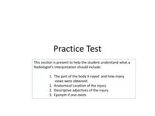
Practice Test This section is present to help the student understand what a Radiologist’s interpretation should include: 1. The part of the body X-rayed and how many views were obtained. 2. Anatomical Location of the injury 2. Descriptive adjectives of the injury 3. Eponym if one exists
Case 1 Your Interpretation = Ask yourself: Body part? How many views? Correct location? Correct descriptive adjectives to characterize the fractures and their positioning. Eponym? Radiologists Interpretation: Left forearm 2 views—AP and Lateral Distal 1/3 radius fracture with minimal displacement and 35oangulation on the lateral view with the apex volarly. Distal 1/3 ulna fracture with complete displacement and shortening less than 1 cm. It also has 35o angulation with the apex volarly.
Case 2 Your Interpretation = Ask yourself: Body part? How many views? Correct location? Correct descriptive adjectives to characterize the fractures and their positioning. Eponym? Radiologist: L elbow. 4 views. There is a nondisplaced intra-articular radial head fx with a anterior and posterior fat pad sign present.
Your Interpretation = Ask yourself: Body part? How many views? Correct location? Correct descriptive adjectives to characterize the fractures and their positioning. Eponym? Case 3 Radiologist: R tib/fib, 2 views, oblique fracture of the proxdiaphysis at the fibular neck, minimally displaced.
Case 4 Your Interpretation = Ask yourself: Body part? How many views? Correct location? Correct descriptive adjectives to characterize the fractures and their positioning. Eponym? Radiologist: Left 2nd finger, distal phalanx. There is a comminuted tuft and shaft fracture with mild to moderate displacement of the distal fragments.