Download
1 / 19
280 likes | 914 Vues
ANTERIOR & MEDIAL COMPARTMENTS OF THIGH. Prof. Ahmed Fathalla Ibrahim Professor of Anatomy College of Medicine King Saud University E-mail: ahmedfathala@hotmail.com. OBJECTIVES. At the end of the lecture, students should: List the name of muscles of anterior compartment of thigh.

E N D
ANTERIOR & MEDIAL COMPARTMENTS OF THIGH Prof. Ahmed FathallaIbrahim Professor of Anatomy College of Medicine King Saud University E-mail: ahmedfathala@hotmail.com
OBJECTIVES At the end of the lecture, students should: • List the name of muscles of anterior compartment of thigh. • Describe the anatomy of muscles of anterior compartment of thigh regarding: origin, insertion, nerve supply and actions. • List the name of muscles of medial compartment of thigh. • Describe the anatomy of muscles of medial compartment of thigh regarding: origin, insertion, nerve supply and actions. • Describe the anatomy of femoral triangle & adductor canalregarding: site, boundaries and contents.
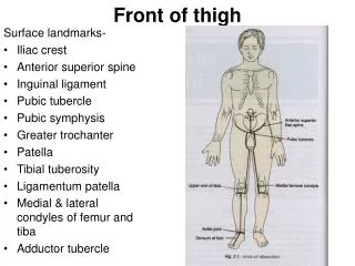
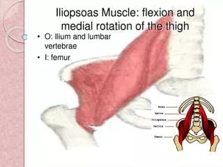
The thigh is divided into 3 compartments by 3 intermuscular septa (extending from deep fascia into femur) • Anterior Compartment • Extensors of knee: • Quadriceps femoris • Flexors of hip: • 1. Sartorius • 2. Pectineus • 3. psoas major • 4. Iliacus • Nerve supply: • Femoral nerve • Medial Compartment • Adductors of hip: • 1. Adductor longus • 2. Adductor brevis • 3. Adductor magnus • (adductor part) • 4. Gracilis • Nerve supply: • Obturator nerve • Posterior Compartment • Flexors of knee & extensors of hip: • Hamstrings • Nerve supply: • Sciatic nerve
ANTERIOR COMPARTMENT OF THIGH I NERVE SUPPLY: Femoral nerve PM P S V L RF Quadriceps femoris VastusIntermedius (deep to rectus femoris) V M
SARTORIUS ORIGIN Anterior superior iliac spine INSERTION Upper part of medial surface of tibia ACTION (TAILOR’S POSITION) Flexion, abduction & lateral rotation of hip joint Flexion of knee joint S S
PECTINEUS ORIGIN: Superior pubic ramus INSERTION: Back of femur (below lesser trochanter) ACTION: Flexion & adduction of hip joint P P
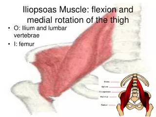
ILIOPSOAS: ILIACUS & PSOAS MAJOR INSERTION: Lesser trochanter of femur I P M I P M ACTION: Flexion of hip joint
QUADRICEPS FEMORIS ORIGIN: Rectus femoris: Anterior inferior iliac spine (Hip bone) Vastusintermedius: Front of shaft of femur Vastusmedialis: Posterior border of femur Vastuslateralis: Posterior border of femur
QUADRICEPS FEMORIS INSERTION: Into PATELLA (Patella is a sesamoid bone) From patella into TUBEROSITY OF TIBIA through LIGAMENTUM PATELLAE (PATELLAR LIGAMENT) ACTION: Extension of knee joint
MEDIAL COMPARTMENT OF THIGH MUSCLES: Adductor longus Adductor brevis Adductor magnus (Adductor part) Gracilis ACTION: ADDUCTION OF HIP JOINT N.B.: Gracilis also flexes knee joint NERVE SUPPLY: OBTURATOR NERVE AB AL G AM Adductor magnus (Adductor portion) Adductor magnus (Hamstring portions)
Origin • Inferior pubic ramus • Ischialramus • Body of pubis • Body of pubis • Inferior pubic ramus Adductor part Adductor hiatus Hamstring part Adductor magnus (adductor part) Gracilis Adductor longus Adductor brevis Insertion • Upper part of medial • surface of tibia • (behind sartorius) • Posterior border of femur (Linea Aspera)
FEMORAL TRIANGLE SITE: Upper third of front of thigh BOUNDARIES: Base: inguinal ligament Lateral: medial border of sartorius Medial: medial border of adductor longus ROOF: Skin Fasciae: superficial & deep FLOOR: From medial to lateral Adductor longus Pectineus Psoas major Iliacus Base: inguinal ligament I PM Lateral P A L Medial S
FEMORAL TRIANGLE CONTENTS: Femoral vein Femoral artery Both vein & artery are enclosed in a fascial envelope (Femoral sheath) Femoral nerve Deep inguinal lymph nodes
DEFINITION: A fascial envelope for femoral artery & vein SITE: In middle third of front of thigh EXTENT: From apex of femoral triangle to adductor hiatus BOUNDARIES: *Roof: Sartorius *Floor: Adductor longus & magnus ADDUCTOR CANAL
SUMMARY MUSCLES OF ANTERIOR COMPARTMENT OF THIGH: • Flexors of hip: Sartorius, pectineus, psoas major & iliacus (all are inserted into femur EXCEPT: Sartorius: inserted into tibia) N.B.: Tailor’s position performed by sartorius: flexion, abduction & lateral rotation of hip + flexion of knee. • Extensors of knee: Quadriceps femoris • All parts originate from femur EXCEPT: Rectus femoris: from hip • All parts are inserted into patella • NERVE SUPPLY: femoral nerve
SUMMARY MUSCLES OF MEDIAL COMPARTMENT OF THIGH: • ACTION: • All muscles adduct hip joint. • Gracilis also flexes knee joint. • ATTACHMENTS: • All muscles originates from pubic bone. • All muscles are inserted into posterior border of femur EXCEPT: gracilis: into tibia (as sartorius) • NERVE SUPPLY: Obturator nerve
QUESTION 1 • Which one of the following muscles is supplied by femoral nerve? • Sartorius • Gracilis • Adductor longus • Adductor brevis
QUESTION 2 • Which one of the following muscles is inserted into the tibia? • Sartorius • Pectineus • Iliacus • Adductor longus