Download
1 / 18
180 likes | 508 Vues
Anterior, Lateral Compartments of the Leg & Dorsum of the Foot. Dr Saeed Vohra. Dr Jamila EL Medany. &. OBJECTIVES At the end of the lecture, student should be able to: Identify the deep fascia of leg Identify the fascial compartments of the leg.

E N D
Anterior, Lateral Compartments of the Leg & Dorsum of the Foot Dr SaeedVohra Dr Jamila EL Medany &
OBJECTIVES At the end of the lecture, student should be able to: • Identify the deep fascia of leg • Identify the fascial compartments of the leg. • Describe the anatomy of the anterior & lateral compartments. • List the contents of each compartment (muscles, vessels & nerves). • Describe the anatomy and contents of the dorsum of the foot
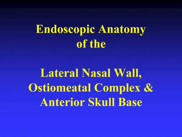
Fascial Compartments of the Leg • The deep fascia surrounds the leg and attached to anterior & medial borders of tibia. • Intermuscular Septa • Two pass from the deep aspect of this fascia to be attached to : • Anterior border of fibula(Anterior fascial septum) • Posterior border offibula(Posterior fascial septum) • Interosseous membrane: • A thin& strong membrane, that binds the interosseous borders of tibia & fibula. It binds the two bones and provides attachment for muscles.
Compartments of the Leg • Together with the interosseus membrane, the septa divide the leg into 3 compartments :1-Anterior • 2-Lateral (Peroneal) • 3-Posterior • Each compartment has its own muscles, blood supply and nerve supply.
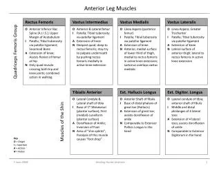
Action Insertion Origin
Extensor Retinacula • Thickening of deep fascia that keep the long tendons around ankle joint in position • Superior Extensor Retinaculum: • Attached to anterior borders of tibia & fibula above ankle • Inferior Extensor Retinaculum: • Y-shaped band located inferior to ankle
Structures passing Deep to Extensor Retinacula From Medial to Lateral: 1.Tibialis anterior 2. Extensor Hallucislongus. 3. Anterior tibialVessels 4. Deep peronealNerve. 5. Extensor Digitorumlongus. 6.Peroneustertius.
Insertion Action Origin
Peroneal Retinacula Superior & Inferior peronealretinacula Connect the lateral malleolus to calcaneum & hold the tendons of peroneuslongus & brevis Synovial Sheaths of Peroneal Longus & Brevis • Tendons of peronei are surrounded by a single common tubular synovial sheath,deep to inferior peroneal retinaculum they have separate sheaths
Deep Fascia of Dorsum of Foot • It is very thin, but just distal to ankle joint, it is • thickened to formInferior extensorretinaculum
Insertion of Long Extensor Tendons • The tendon of Ex dig longusdivides into (4) to the lateral four toes. • Each tendon to the 2nd , 3rd & 4th toes is joined on its lateral side by a tendon of Ex dig brevis. • The extensor tendons form • A fascial Expansion (Extensor Expansion) on the dorsum of each toe. • The expansion divides into (3) parts: • Central:inserted into the base of middle phalanx. • Two lateral : inserted into the base of distal ph. • The EX receives insertion of Interossei & Lumbrical muscles.
Synovial Sheaths of Extensor Tendonson the Dorsum of Foot A separate sheath for each of: Tibialis anterior Extensor hallucislongus A common sheath for : Extensor digitorumlongus & peroneustertius, It extends to the level of base of 5th metatarsal bone.