Download

1 / 37
370 likes | 843 Vues
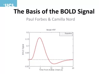
2010. Basis of the BOLD signal. Physics and physiology Louise McDonald and Yen Yu 24 November 2010. Aim. 2010. Explain physics of MRI and fMRI very simply P roton spin Magnets used in the scanner Image formation Contrasts, e.g. T 1 , T 2 , T 2 * Only use essential terminology

E N D
2010 Basis of the BOLD signal Physics and physiology Louise McDonald and Yen Yu 24 November 2010
Aim 2010 • Explain physics of MRI and fMRI very simply • Proton spin • Magnets used in the scanner • Image formation • Contrasts, e.g. T1, T2, T2* • Only use essential terminology • A few numbers • Very few subscripts • NO EQUATIONS!!
History 2010 • 1924 – Pauli suggested that atomic nuclei might spin and therefore have magnetic properties • 1937 – Rabi showed that atomic nuclei (in gases) can absorb energy from magnetic fields = magnetic resonance (MR) • 1945 – Purcell and Bloch demonstrated MR in solids and liquids • 1972-1976 – Lauterbur and Mansfield’s work led to localisation of MR signals in 3D using echoplanar imaging Isidor Rabi Nobel Prize 1944 Edward Purcell and Felix Bloch Nobel Prize 1952 Paul Lauterbur and Peter Mansfield Nobel Prize 2003
History 2010 • 1st MR image of a human body in 1977 • 0.05T, 2 mins/voxel, 4h to get the image
Protons and spin 2010 • Protons • Hydrogen nuclei • Very common in body tissue, which is about 80% water • Protons spin • Usually in random directions • Line up in magnetic fields • Millions of protons in a typical voxel; 100,000 or more voxels in a brain scan Proton spinning (at rest) Direction of magnetic field
Proton spins 2010 • Proton spins are important • At rest, the axes of the spins align with the static magnetic field • After excitation, the axes of the spins precess about the magnetic field lines. (Precession frequency is 64MHz in a 1.5T magnetic field) Direction of magnetic field (B0) Proton spinning (at rest) Direction of magnetic field (B1) Direction of magnetic field (B0)
What happens to protons in an MRI scan 2010 • When protons are at rest, MR signal cannot be detected • In order to detect an MR signal, you first need to excite the protons • The RF transmitter coil generates a magnetic field (B1) at right angles to the static magnetic field (B0) • The protons start to precess around the B1 magnetic field • When the B1 magnetic field is switched off, protons gradually go back to where they were • The relaxing protons emit a signal which can be detected by the RF receiver coil • Spin precession • Following excitation, the frequency of the precession of the proton spins is determined by the strength of the magnetic field • X, Y and Z gradient coils are used to apply gradients of magnetic field • This changes the local spin precession frequency and is used to determine proton spin locations • A typical sequence consists of: excitation-apply gradients-measure signal
Magnets in the scanner 4T magnet 2010 Gradient coils RF receiver coil Gradient coil (inside) Shimming coil Radiofrequency receiver coil Static magnetic field coil
Magnets in the scanner 2010 • Layers of magnetic coils in the scanner • Static magnetic field coil (B0) – lines up proton spins • Shimming coils – make static magnetic field homogeneous • Radiofrequency coils – transmit (i.e. sends the B1 field) and receive MR signal • X, Y, Z gradient coils – localise signal in 3D
Sequence of events in an MRI scan 2010 Static coil is on all the time Shimming coil comes on before the start of scanning and maintains constant settings throughout scanning RF transmitter coil goes on and off during scanning X, Y and Z gradient coils come on in various sequences and combinations (details not shown here) after the RF transmitter coil is switched off and they help to localise the MR signal in 3D RF receiver coil collects MR signal with location information when the RF transmitter coil is off
Static magnetic field 2010 • Very powerful – 1.5 Tesla or more • Earth’s magnetic field = 0.000031 T • Small fridge magnet = 0.005 T • Magnet in a car scrapyard = 1 T
Static magnetic field 2010 • On all the time • Creates a strong (ideally) homogenous magnetic field • Lines up proton spins, which is needed for other coils to work properly
Shimming coil 2010 • MR needs a homogenous magnetic field • Magnetic fields often not homogenous, especially with a human inside them • Can cause artefacts in scan such as image distortions and drop outs • Shimming coil improves this • Shimming is done before the scan and the optimal settings are kept throughout the scan sinuses ear canals
Radiofrequency (RF) coils 2010 • RF coils are used to excite/flip the spins (transmit) and measure signal (receive) • Transmit and receive can be done using the same or different coils. • One coil is located inside the bore (generally used for transmission) and one is located around the subject’s head (normally used to receive signal) RF transmitter coil RF receiver coil
RF transmitter coil (RF transmitter coil is off) • At rest, the axis of the proton’s spin is aligned along B0. • Switching on the RF transmitter coil flips the proton’s spin (flipping of the magnetization). • If it’s a short burst of excitation from the RF transmitter coil, the axis of the proton’s spinprecesses about the RF field B1 • When the axis of the proton’s spin is flipped by 90o, it ends up in the horizontal plane (typical excitation). • With a longer burst of excitation from the RF transmitter coil, the axis of the proton’s spin points down (180o pulse, or inversion) Proton spinning (at rest) Proton’s axis of spin flipped by 90° Direction of magnetic field (B1) from the RF transmitter coil Direction of magnetic field (B0) from the static coil Proton’s axis of spin flipped by 180° 2010
RF receiver coil (RF transmitter coil is switched off) • After protons have been flipped, the RF transmitter coil is turned off • Flipped protons gradually return to their ‘at rest’ spins • Take different amounts of time, depending on the tissue type • RF receiver coil detects this • This is the basis of the MR signal Proton’s axis of spin flipped by 180° Proton’s axis of spin starts to realign with B0 Direction of magnetic field (B0) from the static coil Proton spinning (at rest) 2010
X, Y, Z gradient coils 2010 • RF receivercoil detects the MR signal but location information also needed • X, Y, Z gradient coils locate the MR signal in 3 dimensional space • Make the banging noise during scans
X, Y, Z gradient coils 2010 • Precession frequency is determined by field strength • The field strength can be changed along a gradient in any direction • This creates systematic differences in precession frequency along the directions of the magnetic field gradients • The frequency response from the spins determines their location • Gradient directions: • X is horizontal (e.g. left to right) • Y is vertical • Z is horizontal (e.g. head to toe)
X, Y, Z gradient coils 2010 • Change the strength of the magnetic field along a gradient in different directions X gradient coil increases magnetic field with a gradient of up to 40 mT/m from left to right (in this example) Z gradient coil increases magnetic field with a gradient of up to 40 mT/m from head to toe (in this example) Y gradient coil increases magnetic field with a gradient of up to 40 mT/m from bottom to top (in this example)
Image formation 2010 • MR signals received by the RF coil are processed • Variations in MR signal come from how long it takes proton spins to flip back (relaxation) and whether they have flipped back completely before the next RF transmission pulse • Slices along z-axis
Image formation 2010 • Following excitation, gradient pulses are issued in various sequences and the MR signal is measured • Depending on the sequence in which the gradients are played out, different trajectories can be achieved in each 2D slice: Straight lines = echoplanar imaging Spiral imaging
Image formation 2010 • Results in images of frequencies called k-space Picture of the Mona Lisa in k-space Picture of the Mona Lisa in real space
Image formation 2010 • The signal is acquired in k-space. An image in real space is obtained using Fourier transforms Inverse Fourier transform The same brain image in real space Image of a brain in k-space
Contrasts 2010 • The dynamics of the signal after excitation (T1, T2 relaxations) depend on the tissue properties • White and grey matter have different T1 and T2 relaxation times. As a result, the amount of signal from each tissue type can be tuned, leading to image contrast • The number of protons in each voxel (i.e. proton density) also affects the image contrast
Contrasts 2010 • Types of contrast • T1 – used for static, detailed images of brain • T2 – used for static, detailed images of brain • T2* – used for BOLD • Repetition time (TR), echo time (TE) and excitation flip angle all have an effect on image contrast.
T1 contrast 2010 • Good for anatomically detailed static images of brain • White matter lightest • Grey matter intermediate • Liquid, e.g. CSF, darkest
T1 contrast 2010 • Slower than T2. At 1.5T: • Grey matter 900ms • White matter 600ms • CSF 4000ms • Intermediate repetition time (TR) • Short echo time (TE) to avoid picking up T2 contrast • More sensitive to artefacts than T2 (T1 uses gradient-echo imaging) This subject was wearing a hair band with a ~2 mm copper clamp. Left: with hair band. Right: without. Source: Jorge Jovicich
T2 contrast 2010 • Like a photographic negative of T1 • White matter darkest • Grey matter intermediate • Liquid, e.g. CSF, lightest • Used to image tumours and other pathologies • Related to T2* which is important for BOLD
T2 contrast 2010 • Faster than T1. At 1.5T: • Grey matter 100ms • White matter 80ms • CSF 2000ms • Intermediate echo time (TE) • Long repetition time (TR) to avoid picking up T1 signal • Less sensitive to artefacts than T1 (T2 uses spin-echo imaging)
T2* contrast 2010 • Similar to T2 • Liquid is brightest • Uses gradient-echo imaging • More sensitive to artefacts than T2 • Boring artefacts • Sinuses, ear canals • Interesting artefacts, aka data • Blood flow sinuses T2* image ear canals T2* image superimposed on a T1 image
Recipe for MRI 2010 • 1) Put subject in big magnetic field (leave him there) • 2) Transmit a magnetic field at a 90° angle into subject [about 3 ms] • 3) Turn off 90° magnetic field • 4) Receive MR signal re-transmitted by subject • Manipulate re-transmission with gradients of magnetic fields during this readout interval [10-100 ms] • 5) Store measured MR signal data vs. time • Now go back to 2) to get some more data • 6) Process raw data to reconstruct images • 7) Allow subject to leave scanner (this is optional)
Hold that thought ... 2010 • Main points for understanding BOLD signal • MRI scanners can distinguish between tissue types • e.g. white matter, grey matter, liquid • T2* contrast is used for measuring BOLD signal
Acknowledgments and references 2010 • Antoine for teaching me as much physics as anyone possibly could have • Yen for helping me to get rid of the worst mistakes in my first draft • Previous presenters on Methods for Dummies • Professor Jody Culham of University of Western Ontario for really useful slides and pictures (and to Lea Ernst for the link) http://psychology.uwo.ca/fmri4newbies/Tutorials.html • Huettel SA, Song WA, McCarthy G (2009) Functional Magnetic Resonance Imaging (2nd ed.). Sunderland (Mass.), USA: Sinauer Assoc.
Another summarythat might be useful Magnetic field 2010 Tissue protons align with magnetic field (equilibrium state) RF pulses Protons absorb RF energy (excited state) Spatial encoding using magnetic field gradients Relaxation processes Relaxation processes Protons emit RF energy (return to equilibrium state) MR signal detection Repeat K-space Fourier transform IMAGE
What happens to protons in an MRI scan (the real explanation) 2010 • When protons are at rest, MR signal cannot be detected. In order to detect an MR signal, you first need RF excitation • RF excitation/flipping the magnetization • RF excitation is used to flip the magnetization perpendicular to B0. • When the RF is switched off, protons precess about the B0 field and signal can be detected • Energy emitted by the protons when they precess • Spin precession • Following excitation, precession of the spins is determined by the strength of the magnetic field • Applying gradients of magnetic field changes the local spin precession frequency and is used to determine spin’s locations • A typical sequence consists of: excitation-apply gradients-measure signal