Download
1 / 36
370 likes | 684 Vues
March 25, 2003. The Structural Basis For Calcium Signal Transduction. Walter J. Chazin Center for Structural Biology Vanderbilt University, Nashville TN E-mail: Walter.Chazin@vanderbilt.edu http://structbio.vanderbilt.edu/chazin. Calcium Signal Transduction.

E N D
March 25, 2003 The Structural Basis For Calcium Signal Transduction Walter J. Chazin Center for Structural Biology Vanderbilt University, Nashville TN E-mail: Walter.Chazin@vanderbilt.edu http://structbio.vanderbilt.edu/chazin
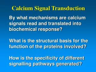
Calcium Signal Transduction By what mechanisms are calcium signals read and translated into biochemical response?What is the structural basis for the function of the proteins involved?How is the specificity of different signalling pathways generated?
Why Go To All The Trouble? Direct, rational and efficient targeting of biological activity with high selectivityAb initio design of biologicalfunction/ activityApplications in Therapeuticsand Biotechnology
Calcium Signal Transduction mM Ca2+ 0.110 mM mM Target
EF-hand Calcium Binding Proteins Function by converting the ionic signal into a biochemical responseCalcium signal transduction involvesa Ca2+ binding induced switch froman “off state” to an “on state”that can interact with target(s)Calmodulin is the paradigm system
Step 1: Calcium-induced Activation Structure Response to Ca2+ binding
Binding of Ca2+ in the EF-hand • Calcium Coordination • 7 oxygen atoms from: • 3 mono-dentate side chains • 1 backbone carbonyl • 1 water H-bonded to side chain • 1 bi-dentate side chain • Specific geometry: pentagonal bi-pyrimid 7 5 9 3 1 12
The Functional Unit: EF-hand DomainNo Isolated EF-hands 2 EF-hands Calmodulin (2 domains) Domain
Generating Clean Signal Readout The change in calcium concentration in the cell has to be small: 50-100 foldThis poses special challenges for the proteins that must have a clean separation between on and off statesCooperative binding of Ca2+ ions is an essential property for signalling What is cooperativity? How is it generated?
The Structural Effect Induced by the Binding of Calcium Ca2+ CaM-N • The structure of the EF-hand domain is changed
Activation of Typical Ca2+ Sensors Accessible Hydrophobic Surface Calmodulin N-terminal Domain
Example: Calmodulin-Mediated Activation of Protein Kinases Myosin light chain kinase
Now we understand how the calcium signal is read and transduced into biochemical response.The next step is to determine how the diversity in the functions of EF-hand proteins is achieved.EF-hand proteins function as both Ca2+ sensors and signal modulators
More Than Just Ca2+ Sensors!!! EF-hand Proteins as Signal Modulators • Shape signal • Buffer • Transport
Structure-Function Relationships EF-hand proteins have homologous sequences and very similar structures, yet diversity in functionHow does nature fine-tune the protein sequence to achieve diversity and specificity of biological action?1. Differences in the response to Ca2+
Ca2+-Induced Conformational Changes Calmodulin (N domain) Calbindin D9K (Signal Modulator) (Sensor)
Functional Diversity and Specificity EF-hand proteins have homologous sequences and very similar structures, yet diversity in functionHow does nature fine-tune the protein sequence to achieve diversity and specificity of biological action?2. Differences in structural organization
S100 Proteins: Unique Architecture • S100 proteins have a unique dimeric structure Potts. et al., 1995
Basic Structural Unit is 4 EF-Hands! • Interdigitated side chains • A single contiguous hydrophobic core
Mode of Action Must Be Unique • S100 proteins have a unique dimeric structure • The mode of signal transduction must be distinct from calmodulin • Smaller changes in conformation Potts. et al., 1995
Ca2+-induced Conformational ChangeS100s Are Different From Calmodulin Ca2+ Sensor S100B CaM-N • S100 protein response is much smaller than typical Ca2+ sensors
Mode of Action Must Be Unique • S100 proteins have a unique dimeric structure • The mode of signal transduction must be distinct from calmodulin • Smaller changes in conformation • Activation must be different from CaM Potts. et al., 1995
The CaM Wrap-around Paradigm Ca2+ Target
MLCK S100s Bind Targets Differently p53 Calmodulin/MLC Kinase S100B/p53 • No wrap-around possible for S100 proteins! • Dimeric structure has 2 symmetric binding sites
S100 Protein Quaternary Structure Dimer Hexamer
Many S100 Proteins Oligomerize S100A9 S100A12 Dimer Tetramer
Functional Diversity and Specificity Diversity from Differences in Quaternary Structure? Tetramer Dimer Hexamer Octamer
Structure-Function Relationships EF-hand proteins have homologous sequences and very similar structures, yet diversity in functionHow does nature fine-tune the protein sequence to achieve diversity and specificity of biological action?3. Differences in target binding
Specificity: Calmodulin vs CentrinDifferences in the Binding Sites for Different Proteins calmodulin centrin • Extremely similar structures, but subtle details different
Structural Basis of Functional Diversity opposite charge extra cleft extra pocket Centrin Calmodulin • Also a critical role for target to match the binding site!
Differences in Hydrophobic Surface Apo S100A6 Apo S100B Ca2+ loaded S100A6 Ca2+ loaded S100B • Differences in D hydrophobic surface induced by Ca2+ binding
Differences in Electrostatic Surface S100B-P53 S100A11-Annexin-I • Complemented by the properties of the target
Functional Diversity and SpecificityDifferent Binding Modes for Different Proteins S100A10/annexin II S100A11/annexin I S100B/p53 S100A9/Chaps
Functional Diversity and SpecificityA New Concept!! S100B-Ndr S100B-p53 • Different binding modes for the same • S100 protein with different targets!!
Summary of Factors Providing Functional Diversity and Specificity • Differences in the architecture and responses to the binding of calcium ions • Sequence variability of residues at the surface alters the character of binding sites • The complementarity of the binding surface and target leads to different binding modes • Different modes for different proteins • Multiple modes for each protein?