Understanding ECG Paper and Standardization in Electrocardiography
This guide explains the importance of standardization signals in ECG paper, emphasizing their role in assigning value to the smallest squares on the graph. It details how different speeds of ECG paper affect time representations and highlights common technical artifacts that can distort readings. The classification of arrhythmias is outlined, identifying normal sinus rhythms and various disturbances in impulse formation. With practical solutions for minimizing interference caused by patient movement or electronic devices, this resource serves as a crucial reference for accurate ECG interpretation.

Understanding ECG Paper and Standardization in Electrocardiography
E N D
Presentation Transcript
ECG Paper and Standardization • Standardization Signal • Without a standardization signal, the ECG paper is merely graph paper. By entering a signal, you place value on each of the smallest squares. • At 1 cm=1 mv each tiny square represents 0.1 mv in height (amplitude) and 0.02 seconds in width (duration) at a paper speed of 50 mm/sec. • At a paper speed of 25 mm/sec, each tiny square represents 0.04 seconds, every large box (5 small boxes) represents 0.20 seconds, and every 15 large boxes (set) = 3 seconds. • The calibration signal can be changed, if necessary, to affect amplitude only.
Baseline or Isoelectric Line Positive charges Negative charges
Common Technical Pitfalls • Incorrect lead placement • Paper speed • Motion artifacts • Breathing/respiratory movement • Purring
Artifacts • The word artifact is similar to artificial in the sense that it is often used to indicate something that is not natural (i.e. man-made). In electrocardiography, an ECG artifact is used to indicate something that is not "heart-made." These include (but are not limited to) electrical interference by outside sources, electrical noise from elsewhere in the body, poor contact, and machine malfunction. Artifacts are extremely common, and knowledge of them is necessary to prevent misinterpretation of a heart's rhythm
Muscle Tremor Interference • If your patient is not calm and comfortable, or just really nervous and shaky…the reading may look like this. Also caused by happy, purring feline patients • Reapplying or readjusting the clips may help • A towel or blanket can be placed on patient to help calm them • You can also place your hand on the chest of the patient, taking care not to apply too much pressure (will interfere with reading)
Electrical Interference • Remember that rubber mat and how you checked your machine for causes of bad ground? • Interference can be caused by other machinery such as a pulse oximeter or BP monitor that is hooked up to animal; even fluorescent lighting
ECG Tracings of Common Arrhythmias • Please refer to your ECG handout • Reading assignment VTDRG: Pg 205-220, 338-345
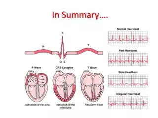
Classification of Arrhythmias • Definition • An abnormality in the rate, regularity, or site of origin of the cardiac impulse • A disturbance in conduction of the impulse such that the normal sequence of activation of the atria and ventricles is altered
Classification of Arrhythmias • Normal sinus impulse formation • Normal sinus rhythm • Sinus arrhythmia • Disturbances of sinus impulse formation • Sinus bradycardia • Sinus tachycardia • Disturbances of supraventricular impulse formation • Atrial premature complexes • Atrial tachycardia • Atrial flutter • Atrial fibrillation
Disturbances of ventricular impulse formation • Ventricular premature complexes • Ventricular tachycardia • Ventricular systole • Ventricular asystole • Ventricular fibrillation Disturbances of impulse conduction • Sinus arrest or block • Atrial standstill • Ventricular pre-excitation • First-degree AV block • Second degree AV block • Third degree AV block • Left bundle branch block • Right bundle branch block
Building Blocks for Arrythymia Interpretation • Recognize the site of origin of the abnormal beat • Recognize deviations from the normal rate of automaticity for that site • Site of Origin • Atrial • Positive deflection P waves are present with a constant P-R interval and normal duration QRS complex • Junctional • Negative deflection P waves, or no P waves with a normally conducted short QRS complex • Ventricular • No P waves are evident, QRS complexes are wide and bizarre appearing and may be positive or negative polarity depending on which ventricle is the site of origin