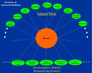
Microbiology
Microbiology. Definition: Microbiology is the science that deals with microorganisms that can not be seen by the naked eye . The Importance of Microbiology : 1-The Environment : Microbes are responsible for the cycling of carbon, nitrogen and phosphorus .

Microbiology
E N D
Presentation Transcript
Microbiology • Definition: • Microbiology is the science that deals with microorganisms that can not be seen by the naked eye. The Importance of Microbiology: 1-The Environment: Microbes are responsible for the cycling of carbon, nitrogen and phosphorus. 2- Medicine:Production of antibiotics. 3- Food:Wine and bread making, cheese and milk products production. 4- Biotechnology: Some microorganisms have been used to synthesize many chemicals such as acetone and acetic acid. 5- Research: Microbes have been used as model organisms for the biochemical and medical investigations. Dr. Rania Alhady
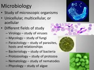
Microbiology What is a Microbe? A microbe or microorganism is: A- A living creature which can not be seen except by microscope. B- It is simple in structure. C- Usually unicellular. D- It can be either prokaryotic cell or a eukaryotic cell. Dr. Rania Alhady
Microbes Prokaryotes and Eukaryotes: Dr. Rania Alhady
Microscopy Microscopy: It is the technical field of using a microscope to view samples and objects that can not be seen by unaided eye ( objects that aren't within the resolution range of normal eye). A microscope is the most important piece of equipment used in clinical microbiology. Microscopy forms 70-90% of the work. Dr. Rania Alhady
Microscopy Working principle of a microscope: A microscope is a magnifying instrument. The magnified image of the object (specimen) is first produced by a lens close to the object called the objective lens. Objective lens magnification = 4 X to 100 X A second lens near the eye called the eyepiece enlarges the primary image, converting it into one that can enter the pupil of the eye. Eyepiece magnification = 8 X to 12 X typically. (but 10 X is most common) Total magnification = objective magnification* eyepiece magnification (4, 10, 40, 100 X) * ( 10 X) So, final magnification ranges from 40 X up to 1000 X Dr. Rania Alhady
Microscopy Resolution of Microscope: It is the ability to distinguish two very small and very closed objects as separate entities. Resolution is best when the distance separating the two tiny objects is small. So, Resolution can be defined in a different way as: It is the smallest distance that separate two sources of light points reflected from two particles close together on object. Resolution is determined by certain physical parameters that include the wavelength of light, and the light-gathering power of the objective & condenser lenses. Resolution = light wavelength / 2 Example:yellow light of a wavelength of 0.4 μm give a resolution of 0.2 μm. Dr. Rania Alhady
Microscopy Types of microscopy: 1- The light microscope: (simple and compound). A- The student (Brightfield) microscope B- Phase contrast microscope. C- Darkfield illumination microscope. 2- The Fluorescence microscope. 3- The Electron microscope. 1- The Light Microscope: - Brieghtfield microscope is most likely found in classrooms or labs. - Better equipped classrooms and labs have darkfield&/or phase contrast microscope. Dr. Rania Alhady
Microscopy A-The student microscope: Image quality is based largely on your ability to use the microscope properly. Uses:Brightfieldillumination microscope is mainly used to examine stained smear Or naturally pigmented specimens. Dr. Rania Alhady
Microscopy Light path consists of: Transillumination light source, commonly a halogen lamp in the microscope stand. Condenser lens which focuses light from the light source onto the sample. Objective lens which collects the light from the sample and magnifies the image. Oculars and /or a camera to view the sample image. Objects seen in the light path because: -Natural pigmentations or stains absorb light differentially. Or - They are thick enough to absorb a significant amount of light despite being colorless. Dr. Rania Alhady
Microscopy B-Phase contrast microscope: This is needed to visualize transparentmicroorganisms suspended in a fluid. (does not require staining to view the slide) The phase microscope takes advantage of the fact that light waves passing through transparent slide or objects (cells) emerge in different phases (different times). A special optical system converts difference in phase into difference in intensity. So that some structure appear darker than others. This microscope made it possible to study cell cycle. Dr. Rania Alhady
Microscopy C-Dark field illumination microscope: This method is used for visualizing organisms suspended in fluid and can not be stained by Gram’s stain. Both the structure and the motility of the organisms can be seen. Used for: Unstained biological samples such as a smear from a tissue culture. In this method: the light enters the special condenser which has a central blacked-out area, so that light can not pass directly through it to enter the objective. Instead, the light is reflected to pass through the outer rim of the condenser at a wide angle which illuminates the microorganism by a ring of light surrounding them. Dr. Rania Alhady
Microscopy Micrcoccus species examined by phase contrast microscope ( left) and Leptospira examined by Darkfield microscope (Right): Dr. Rania Alhady
Microscopy Dr. Rania Alhady 2- The Fluorescence Microscope: In this method, microorganisms must be stained with a fluorescent dye such as rhodamine. A fluorescent lamp emits visible light which is filtered off using optical filters. (Ultraviolet (UV) wavelengths will not be filtered off). A fluorescent dye will absorb this UV-light and change it to visible (yellow or orange) light.
Microscopy 3-The Electron Microscope: - Electron microscope is an instrument that magnify very small objects which can not be seen by light microscopy. - Resolution of this microscope is higher than the light one. - The energy source of this kind is the electron beam while light waves are used in the light microscope. - Electromagnetic lenses are used instead of the glass lenses of light microscope. - Visible light of 500 nm wavelength give a resolution of 250 nm. Electron beam of 0.001 nm wavelength give a resolution of 0.0005 nm. Viruses with a diameter of 0.01-0.2 micrometer can be easily seen. Magnification of electron microscope is up to about 10.000.000 X . Dr. Rania Alhady
Microscopy Dr. Rania Alhady
Microscopy Light waves and Electron beam: Dr. Rania Alhady