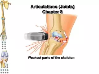
Chapter 9 Joints
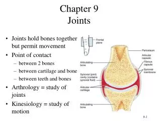
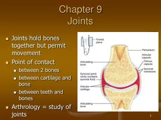
Chapter 9 Joints. Joints hold bones together but permit movement Point of contact between 2 bones between cartilage and bone between teeth and bones Arthrology = study of joints Kinesiology = study of motion. Classification of Joints . Structural classification based upon:

Chapter 9 Joints
E N D
Presentation Transcript
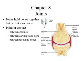
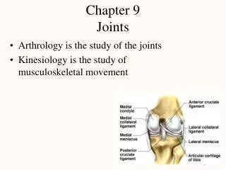
Chapter 9Joints • Joints hold bones together but permit movement • Point of contact • between 2 bones • between cartilage and bone • between teeth and bones • Arthrology = study of joints • Kinesiology = study of motion
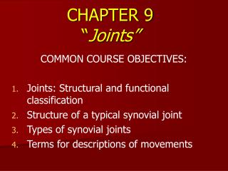
Classification of Joints • Structural classification based upon: • presence of space between bones • type of connective tissue holding bones together • collagen fibers • cartilage • joint capsule & accessory ligaments • Functional classification based upon movement: • immovable = synarthrosis • slightly movable = amphiarthrosis • freely movable = diarthrosis
Fibrous Joints • Lack a synovial cavity • Bones held closely together by fibrous connective tissue • Little or no movement (synarthroses or amphiarthroses) • structural type • sutures • syndesmoses • gomphoses
Sutures • Thin layer of dense fibrous connective tissue unites bones of the skull • Immovable (synarthrosis) • If fuse completely in adults is synostosis
Syndesmosis • Fibrous joint • bones united by ligament • Slightly movable (amphiarthrosis) • Anterior tibiofibular joint and Interosseous membrane
Gomphosis • Ligament holds cone-shaped peg in bony socket • Immovable (amphiarthrosis) • Teeth in alveolar processes
Cartilaginous Joints • Lacks a synovial cavity • Allows little or no movement • Bones tightly connected by fibrocartilage or hyaline cartilage • 2 types • synchondroses • symphyses
Synchondrosis • Connecting material is hyaline cartilage • Immovable (synarthrosis) • Epiphyseal plate or joints between ribs and sternum
Symphysis • Fibrocartilage is connecting material • Slightly movable (amphiarthroses) • Intervertebral discs and pubic symphysis
Synovial Joints • Synovial cavity separates articulating bones • Freely moveable (diarthroses) • Articular cartilage • reduces friction • absorbs shock • Articular capsule • surrounds joint • thickenings in fibrouscapsule called ligaments • Synovial membrane • inner lining of capsule • secretes synovial fluid containing hyaluronic acid slippery) • brings nutrients to articular cartilage
Example of Synovial Joint • Joint space is synovial joint cavity • Articular cartilage covering ends of bones • Articular capsule
Other Special Features • Accessory ligaments • extracapsular ligaments • outside joint capsule • intracapsular ligaments • within capsule • Articular discs or menisci • attached around edges to capsule • allow 2 bones of different shape to fit tightly • increase stability of knee - torn cartilage • Bursae = saclike structures between structures • skin/bone or tendon/bone or ligament/bone
Arthroscopy & Arthroplasty • Arthroscopy = examination of joint • instrument size of pencil • remove torn knee cartilages & repair ligaments • small incision only • Arthroplasty = replacement of joints • total hip replaces acetabulum & head of femur • plastic socket & metal head • knee replacement common
Torn Cartilage and Arthroscopy • Damage to menisci of the knee joint • Visualization of the inside of a joint • arthroscope • requires only small incisions • Repair may include removal of torn cartilage
Nerve and Blood Supply • Nerves to joints are branches of nerves to nearby muscles • Joint capsule and ligaments contain pain fibers and sensory receptors • Blood supply to the structures of a joint are branches from nearby structures • supply nutrients to all joint tissues except the articular cartilage which is supplied from the synovial fluid
Sprain versus Strain • Sprain • twisting of joint that stretches or tears ligaments • no dislocation of the bones • may damage nearby blood vessels, muscles or tendons • swelling & hemorrhage from blood vessels • ankle if frequently sprained • Strain • less serious injury • overstretched or partially torn muscle
Planar Joint • Bone surfaces are flat or slightly curved • Side to side movement only • Rotation prevented by ligaments • Examples • intercarpal or intertarsal joints • sternoclavicular joint • vertebrocostal joints
Hinge Joint • Convex surface of one bones fits into concave surface of 2nd bone • Uniaxial like a door hinge • Examples • Knee, elbow, ankle, interphalangeal joints • Movements produced • flexion = decreasing the joint angle • extension = increasing the angle • hyperextension = opening the joint beyond the anatomical position
Pivot Joint • Rounded surface of bone articulates with ring formed by 2nd bone & ligament • Monoaxial since it allows only rotation around longitudinal axis • Examples • Proximal radioulnar joint • supination • pronation • Atlanto-axial joint • turning head side to side “no”
Condyloid or Ellipsoidal Joint • Oval-shaped projection fits into oval depression • Biaxial = flex/extend or abduct/adduct is possible • Examples • wrist and metacarpophalangeal joints for digits 2 to 5
Abduction and Adduction Condyloid joints Ball and Socket joints
Saddle Joint • One bone saddled-shaped; other bone fits as a person would sitting in that saddle • Biaxial • Circumduction allows tip of thumb travel in circle • Opposition allows tip of thumb to touch tip of other fingers • Example • trapezium of carpus and metacarpal of the thumb
Ball and Socket Joint • Ball fitting into a cuplike depression • Multiaxial • flexion/extension • abduction/adduction • rotation • Examples • shoulder joint • hip joint
Bursae and Tendon Sheaths • Bursae • fluid-filled saclike extensions of the joint capsule • reduce friction between moving structures • skin rubs over bone • tendon rubs over bone • Tendon sheaths • tubelike bursae that wrap around tendons at wrist and ankle where many tendons come together in a confined space • Bursitis • chronic inflammation of a bursa
Summary of Movements at Synovial Joints • Gliding • no change in angle of joint • Angular movements • increase or decrease in angle between articulating bones • flexion, extension, hyperextension • adduction, abduction • circumduction is a combination of above movements • Rotation • bone revolves around its own axis • Special movements • uniquely named movements for jaw, hand and foot
Circumduction • Movement of a distal end of a body part in a circle • Combination of flexion, extension, adduction and abduction • Occurs at ball and socket, saddle and condyloid joints
Rotation • Bone revolves around its own longitudinal axis • medial rotation is turning of anterior surface in towards the midline • lateral rotation is turning of anterior surface away from the midline • At ball & socket and pivot type joints
Special Movements of Mandible • Elevation = upward • Depression = downward • Protraction = forward • Retraction = backward
Special Hand & Foot Movements • Inversion • Eversion • Dorsiflexion • Plantarflexion • Pronation • Supination
Shoulder Joint • Head of humerus and glenoid cavity of scapula • Ball and socket • All types of movement
Glenohumeral (Shoulder) Joint • Articular capsule from glenoid cavity to anatomical neck • Glenoid labrum deepens socket • Many nearby bursa (subacromial)
Supporting Structures at Shoulder • Associated ligaments strengthen joint capsule • Transverse humeral ligament holds biceps tendon in place
Rotator Cuff Muscles • Attach humerus to scapula • Encircle the joint supporting the capsule • Hold head of humerus in socket
Elbow Joint • Hinge joint • trochlea notch of ulna and trochlea of humerus • flexion and extension of elbow • Pivot joint • head of radius and capitulum of humerus • supination and pronation of forearm
Articular Capsule of the Elbow Joint • Radial annular ligament hold head of radius in place • Collateral ligaments maintain integrity of joint
Hip Joint • Head of femur and acetabulum of hip bone • Ball and socket type of joint • All types of movement possible
Hip Joint Structures • Acetabular labrum • Ligament of the head of the femur • Articular capsule
Hip Joint Capsule • Dense, strong capsule reinforced by ligaments • iliofemoral ligament • ischiofemoral ligament • pubofemoral ligament • One of strongest structures in the body
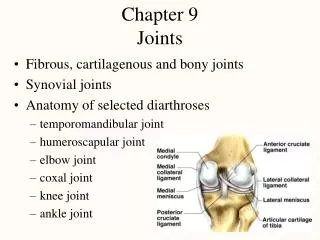
Tibiofemoral Joint • Between femur, tibia and patella • Hinge joint between tibia and femur • Gliding joint between patella and femur • Flexion, extension, and slight rotation of tibia on femur when knee is flexed
Tibiofemoral Joint • Articular capsule • mostly ligs & tendons • Lateral & medial menisci = articular discs • Many bursa • Vulnerable joint • Knee injuries damage ligaments & tendons since bones do not fit together well
External Views of Knee Joint • Patella is part of joint capsule anteriorly • Rest of articular capsule is extracapsular ligaments • Fibular and tibial collateral ligaments
Intracapsular Structures of Knee • Medial meniscus • C-shaped fibrocartilage • Lateral meniscus • nearly circular • Posterior cruciate ligament • Anterior cruciate ligament
Temporomandibular Joint • Synovial joint • Articular disc • Gliding above disc • Hinge below disc • Movements • depression • elevation • protraction • retraction
Atlanto-occipital joints • Atlas and occipital condyles • Condyloid Joint • Flexion • Extension • Slight lateral tilting
Intervertebral Joints • Between bodies and intervertebral discs • symphysis • Between vertebral articular processes • synovial • Flexion • Extension • Lateral flexion
Elbow Joint • Trochlea of humerus, trochlear notch of ulna & head of radius • Pivot and hinge types • Flexion, extension, pronation & supination
Radiocarpal Joint • Articular disc • Condyloid type • Flexion, extension, abduction & adduction
Talocrural Joint • Tibia & fibula with talus • Hinge • Inversion, eversion, plantarflexion & dorsiflexion • Strong joint, seldom dislocates