Classification, Microscopy, Staining
nothing. Classification, Microscopy, Staining. Prokaryotic Classification and Identification. Traditional classification was by Gram stain cellular morphology, arrangement of cells, colony morphology, biochemical characteristics

Classification, Microscopy, Staining
E N D
Presentation Transcript
nothing Classification, Microscopy, Staining
Prokaryotic Classification and Identification • Traditional classification was by Gram stain cellular morphology, arrangement of cells, colony morphology, biochemical characteristics • Modern classification uses DNA sequencing to determine relatedness
Prokaryotic Classification and Identification • 3 methods are used to identify unknown microorganisms in BIO 203
1. Cell morphology and arrangement • Determined by microscopic analysis • Cellular Morphology • 3 basic shapes • Other shapes
Coccus Typical prokaryotic morphologies Spirillum Coccobacillus Spirochete Bacillus Pleomorphic Vibrio
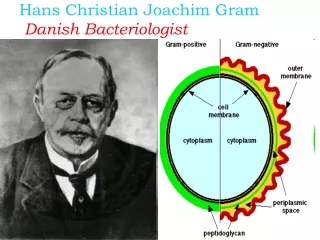
1. Cell morphology and arrangement:Use of Gram Stain • Staining technique which differentiates between 2 large groups of microorganisms • Gram + • Gram – • The staining results are different due to the composition of the cell wall
2. Colony morphology • Macroscopic examination of how the organism grows on different media
3. Biochemical Testing • This means that not all bacteria are created equally • Ex. Not all bacteria can metabolize lactose
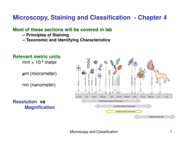
Three factors that determine the quality of an optical image: • Magnification • Resolution • Contrast
Magnification Compound microscope is a 2 lens system: • Objective lens (in the nosepiece) • Ocular lens or eyepiece Total magnification = (magnification of objective lens) X (magnification of ocular lens)
Figure 4.2 Light refraction and image magnification by a convex glass lens-overview Light Glass Air Focal point Inverted, reversed, and enlarged image Convex lens Specimen
Resolution • Is the ability to separate 2 objects that are close together so that they are seen as 2 objects
Figure 4.5 The effect of immersion oil on resolution-overview Microscope objective Microscope objective Lenses Refracted light rays lost to lens More light enters lens Immersion oil Glass cover slip Glass cover slip Slide Slide Light source Specimen Light source With immersion oil Without immersion oil
Contrast • Differences in intensity between two objects, or between an object and background • Most microbes appear colorless against a colorless background = hard to see! • Methods used to increase contrast: Staining • Direct staining
Microscopic Techniques: Dyes and Staining • Purpose: Increase contrast and resolution between microbe and background: makes microbe more visible • Microorganisms are mostly transparent on a slide
Types of Staining • Simple Stains • One stain or dye used to provide contrast • Differential Stains • 2 or more stains/dyes used to provide contrast • Special Stains • Staining cell structures or background
Simple stain Using a single dye to stain cells Examples include: Methylene blue Crystal violet Safranin
Differential stains • Staining procedure to distinguish one group of bacteria from another • Gram stain • Acid Fast • Endospore
Gram Stain Gram + (Purple) Gram – (Pink)
Acid-fast stain • Used to stain organisms of the genus Mycobacterium, which do not readily take up dyes/stains • Contain waxy mycolic acid in cell walls which resists staining
Endospore stain • Endospore stain: used to stain cells which form endospores
Capsule stain • Capsule stain: used to stain cellular capsules
Flagella Stain Flagella • Flagella stain: used to stain the flagella of cells, which is normally too thin to see
Types of Microscopy • Light Microscope • Bright-field (most common, easy to use) • One of most important tools for studying Livingmicroorganisms • Up to 1000x • Electron • Magnify in excess of 100,000x • Fine details of cell surfaces, internal and external structure
Types of Microscopy: Electron • TEM: transmission electron microscope • High resolution • Internal structures of cells • SEM: scanning electron microscope • High resolution images • Surface structures and details of the cell surface