CHAPTER 3 Observing Organisms Through a Microscope
CHAPTER 3 Observing Organisms Through a Microscope. Units of Measurements Microscopy: The Instruments Preparation of Specimens . We will be looking at very small things…. Compound Light Microscope. Compound Light Microscope. Total Magnification Objective lens power x ocular lens power

CHAPTER 3 Observing Organisms Through a Microscope
E N D
Presentation Transcript
CHAPTER 3Observing Organisms Through a Microscope Units of Measurements Microscopy: The Instruments Preparation of Specimens
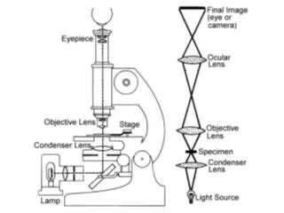
Compound Light Microscope • Total Magnification • Objective lens power x ocular lens power • Resolution • Ability of the lenses to distinguish fine detail • Resolution power of 0.2 μm, • Distinguish 2 points 0.2 μm apart • Refractive index • Change by staining specimens • Two different mediums • Rays change more directions • Oil Immersion • Same index as glass • Improves resolution
STAINS • Salts composed of a positive and a negative ion • One of which is colored (chromophore) • Basic Dyes: positive ion • Attracted to negatively charged bacteria cell • Ex: Crystal violet, methylene blue, malachite green and safranin • Acidic Dyes: negative ion • Stain colors the background surface • Observing overall cell shape, size and capsule • Ex: fuchsin, nigrosin • Called Negative Staining • Three types of staining techniques • Simple, differential and special
SIMPLE STAIN • Aqueous or alcohol sol’n of a single basic dye • Highlight the entire microorganism • Applied to fixed smear for length of time, washed, dried • Mordant (used to intensify) may be added • Increases affinity of a stain • Coat a structure to make it thicker and easier to see (flagella) Examples: methylene blue*, crystal violet, safranin
Staining • Fixed: kills/attaches org. to slide • Thin film spread over slide (smear) and allowed to dry • Pass through flame of Bunsen burner several times or cover with methyl alcohol for 1 min. • Stain is applied and washed with water • Blot with absorbent paper
Differential Stains • React differently with different kinds of bacteria • Used to distinguish • Gram Stain: one of most important staining techniques • 1st used in 1884 by Hans Christian Gram • Searching for method to see cocci in lung tissue • Series of staining and decolorization steps • According to cell wall composition • Gram-positive bacteria have cell walls that contain thick layers of peptidoglycan (90% of wall) PURPLE • Gram-negative bacteria have walls with thin layer of peptidoglycan (10% of wall) and high lipid content PINK
Differential Stains • Gram Stain: • Heat-fixed smear covered with basic purple dye • primary stain (crystal violet) 2. Washed and covered with iodine (mordant), washed off 3. Washed with alcohol (decolorizing agent). Removes purple from the cells of some spp 4. Alcohol is rinsed and slide stained with safranin (basic red dye) 5. Smear washed, blotted dry and examined
GRAM POSITIVE Purple dye and iodine combine in cytoplasm and color it dark violet Thicker peptidoglycan cell wall Traps CV-I inside cell GRAM NEGATIVE Lose the dark violet after decolorization Safranin applied to turn bacteria pink Layer of lipopolysaccharide Alcohol disrupts outer lipopolysaccharide layer and CV-I complex washes out Staphylococcus epidermidis E. coli
http://www.microbelibrary.org/microbelibrary/files/ccImages/Articleimages/keen/Gramstainkeen.htmhttp://www.microbelibrary.org/microbelibrary/files/ccImages/Articleimages/keen/Gramstainkeen.htm
Special Stains Used to color and isolate specific parts Endospore of Bacillus thuringiensis Capsule of Klebsiella pneumoniaeare Flagella of Salmonella
Aseptic Technique • Method that prevents the introduction of unwanted organisms into an environment
Wear appropriate protective equipment • Disinfect working area before you start • Gather all necessary materials • Do any labeling • Never light burner while wearing gloves • Properly adjust the flame of the bunsen burner (small blue cone, not a large plume, nor is it orange) • Once you have flamed your loop DO NOT lay it down or touch it to any surface • Allow loop to cool before you pick up organisms • Ensure you are transferring the correct organisms • Always keep caps and tops in your hand. • Always tape inoculated plates together and incubate them upside down • Discard contaminated materials properly, return supplies to proper storage locations and clean up mess • Disinfect work area when done
Flaming loop • Heat from the base of the wire first and slowly move towards the loop (tip). Heat the wire until it is red hot • The metal must glow orange-red before sterilization is considered complete
Test tube • Remove caps from liquid specimens and replace the caps of the test tubes with the same hand that holds the loop. The caps must be held during the entire procedure and not placed on the desktop • Flame the neck of the tube. Remove a loop full of the culture with the cooled loop and briefly flame the opening of the tube again
Petri Dish • Flame the loop and then open the petri dish just enough to allow the entry of the loop.
Put in Biohazard Bag • Contaminated gloves, paper towels, petri plates, swabs, other non-sharp items
Spilled Culture Material • Clean by saturating with alcohol and wiping with paper towels (dispose of in BioHazard)
WORKS CITED • http://biology.clc.uc.edu/fankhauser/Labs/Microbiology/Bacterial_Smear_&_Staining/06_fix_specimen_P1092682.JPG • http://www.biosynth.com/media/verschiedene/dyes1.JPG • http://www.bigroom.org/images/Sally_MB.jpg • http://student.ccbcmd.edu/courses/bio141/labmanua/lab12/diseases/uti/images/gnrod.jpg • http://people.uleth.ca/~selibl/Biol3200/Morphology04/Btendo.jpg • http://bioinfo.bact.wisc.edu/themicrobialworld/S.typhi.Fla.jpg