
Chapter 2 Epithelium
E N D
Presentation Transcript
Chapter 2 Epithelium Teached by:Xu Fu-Cui
Purpose and requirements 1. Master common features of epithelium. 2. Master distribution & classification of covering epithelium. 3. Master specializations of epithelium. 4. Individual study glandular epi. and glands.
Tissuesare aggregates or groups of cells organized to perform one or more specific funtions. Epithelial tissue (Epithelium) Connective tissue Muscular tissue Nervous tissue classification
1、Characteristics of Epithelium 1). Construction features more cells(arranged tightly),less intercellular substance.
2)Polarity: ---free surface: towards the air or the cavity ---basal surface: face underlying CT, have basement membrane ----lateralsurface:between the adjacent cells 3) No blood vessels, rich in nerve endings
4)location: • Cover the surface of body,line its cavities and tubule or glands
5)Functions • protection: physical / chemical / micro-organism • secretion: mainly for glandular epi. • absorption:small intestine / renal tubule • excretion:sweat gland / renal tubule
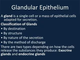
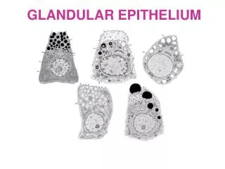
2.TYPES OF EPITHELIA 1)Covering epithelium: the epithelium which cover body surface or line the inner surface of body cavities, tubes and sac,main function are pretection and obsorbtion 2)Glandular epithelium: the epithelium which main function is secretion.
3.Covering epithelium: Principles of classification: • The number of cell layer • Morphologic features of the cell in the surface layer Simple epi.: ---simple squamous epi. ---simple cuboidal epi. ---simple columnar epi. ---pseudostratified ciliated columnar epi. Stratified epi.: ---stratified squamous epi. ---stratified columnar epi. ---transitional epi.
Simple epi. Stratified epi. Stratified epi. Simple epi.
The boundaries between cells are frequently indistinguishable in LM,and the nuclear form often corresponds roughly to the cell shape, so the nuclear is a clue to the shape and number of cells. Shape of cell features of nucleus Squamous flattend cuboidal spherical centrally located Columnar ovoid basally located
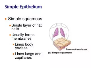
1)simple squamous epi: ---structural feature: /one layer flattened cells, with flattened nucleus
Location:Mesothelium: lining the body cavities (thoracic / pericardial / abdominal cavity)Endothelium: lining the inner surface of the cardiovascular & lymphatic systemOthers: Bowman’s capsule; alveolar; function: transport of materials
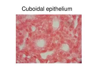
2)simple cuboidal epi.: ---structural feature: • one layer of cells, with same height and width (in side view). • spherical centrally-located nucleus
---distribution: / thyroid /renal tubule ---main function: /Absorption, secretion
3)simple columnar epi. ---structural features: • one layer of columnar cells,with basally located ovoid nucleus • Special structure:striated border(small intestine
goblet cell( small or large intestine): scattered, secreting granules-mucus Location: gastrointestinal tract; gall bladder; uterus ---function: secretion and absorption
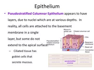
ciliated columnar c. fusiform cell goblet cell basal cell • Foue types of cell, Nu. at different levels 4)pseudostratified ciliated columnar epi.: --- 3 layers of nuclei giving a stratified appearance,but in fact cells vary in height and not all of them reach the surface,they rest on the Basal lamina
Cilia---the surface of columnar cell(clean) • Goblet cells are also present • Basement membrane --- semipermeable membrane , is obviously seen • --distribution:trachea & bronchi & nasal & epididymis. Major function: Secretion, pretection
5)stratified squamous epi.: ---structural features: • deepest(basal)cells: one layer of cuboidal cells • intermediate regions:several layers of polygonal – shaped cells • to the surface: more and more flattened
---distributon: • nonkeratinised:mouth,pharynx, esophagus, urethra and vagina • keratinised: skin Major function: Barrier, protection
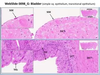
6)transitional epi. ---structural feature: • The layers / shape of cells are flexible according to the degreen of distention of bladder • distended state: 2-3 layers, more flat. • contracted state : 6-7 layers. surface cells are very large and cuboidal in shape, • ---distribution: bladder
4. Epithelial specializations • Free surface • Lateral surface • Basle surface
1)Specialisations of free surface microvilli cilia
①microvilli: ---definition: finger-liked projections of cell-membrane and cytoplasm protruding from the free surface
---structure: • 0.1um in diameter • surface: cell membrane with cell coat • core: microfilament-fixed on terminal web (terminal web: made up of transverse-arranged filament at the apical side of cells)
Function: increase the surface areas Distribution: striated border(LM)----- intestinal epi.cell brush border(LM)------proximal renal tubule A B C
②cilia: ---definition: elongated, mobile projections of cell membrane and cytoplasm protruding from free surface
---structure: • 5-10um long, 0.2um in diameter • surface: cell membrane • core: microtubules(central—a pair of isolated microtubules surrounded by 9 pairs microtubules )
Function:rapid back-and –forth movement, moving fluid and particles in one dirrction over the EP Distribution: respiratory tract A B
2)specializations of the lateral surface EP cells are extremely cohesive , strong mechanical forces to separate ---intercellular junctions-----adhesion、 prevent、providea mechanism for comunication between adjacent cells.
Present in order from apex towards the base of the cell Tight junction (zonula occludens) Intermediate junction (zonula adherens) Gap junction (communicating junction) Desmosome (macula adherens)
structure function tight membrane fusionin close off spacejunction prevent intermediate 15-20nm gap,junction electron-density filament adherens attached microfilament keep shapedesmosom 20-30nm gapattachment plaque firmly connection attached intermediate filamentgap junctionconnexons communication 6-subunits of proteins -2nm channel-hydrophilic
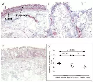
①basement membrane: ---definition: a sheet of membrane-liked material interposed between epi.cells and underlying CT. ---structure: • HE: pink colour wave line
Lamina lucida Basal lamina • EM: three layers --lamina lucida: electron-lucent --basal lamina: electron-dense, produced by epi. Cell --reticular lamina:CT+reticular fibers, produced by CT reticular lamina
---function: • support, connection, fixaton • semi-premeable membrane • induce the movement, proliferation and differentiation of epi.cell
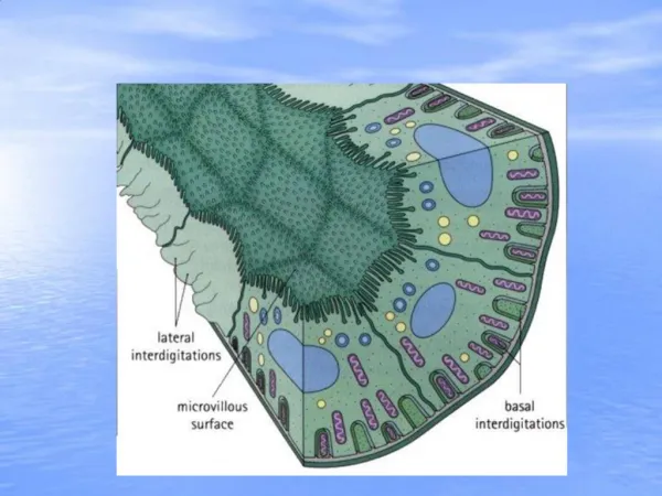
②plasma membrane infolding infolding of cell-membrane with many mitochondria at the basal surface of epi.cell ---function: • increase the basal surface areas • facilitate the passage of water and ions ---distribution: mainly in proximal and distal renal tubule.
Summary What is epithelium? Functions of Epithelium Characteristics of Epithelium Classification of Epithelium Specialization of Epithelium