Download
1 / 16
590 likes | 3.15k Vues
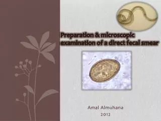
Preparation & microscopic examination of a direct fecal smear. Amal Almuhana 2012. Introduction . Direct fecal smears Direct fecal smears can be used as a quick screening test to check for any intestinal parasite. Advantages &Disadvantages . Advantages:-

E N D
Preparation & microscopic examination of a direct fecal smear AmalAlmuhana 2012
Introduction Direct fecal smears • Direct fecal smears can be used as a quick screening test to check for any intestinal parasite.
Advantages &Disadvantages • Advantages:- • Useful for detecting motile organisms. • Protozoa are often detected via a direct fecal smear. • Quick process.
2) Disadvantages • Small size of the sample limits its usefulness. • You may get inaccurate results. • If your examination finds no evidence of a parasite but the patient actually harbors the parasite, then the results are called a false negative result. False negative results are common with direct fecal smears.
Fecal collection • When collecting - Fecal sample from animal in question. - Relatively fresh - May preserve in refrigerator if exam not immediate. - Free from debris • Storage • Plastic or glass jar/ vial • Plastic cup • Plastic bag
Preparing direct fecal smears(procedure):- • Put small amount of feces on glass slide. • Mix with drop of saline. • Place cover slip on mixtures. • Observe under microscope. Note:- If the feces is already in a liquid state because the animal has diarrhea, obviously no fluid is needed to spread the feces over the slide.
feces+ saline on the slide Mix until it is dispersed Examine under microscope
Direct fecal smear cont. • Direct fecal smears may be examined as : • Awet mount, or can be • Dried and stained.
Wet mount technique • The fecal smear may be examined in its wet state by simply placing a cover slip over the drop of wet fecal material. • This method is most useful looking for trophozoiteswhich can be observed by their characteristic movement and appearance.
Staining • A drop of stain can be added before the cover slip is placed or after having examined the unstained preparation. • The stain will diffuse and then you can examine it. • The iodine will stain the organisms a dark orange brown color. • If you use new methylene blue instead, you will see organisms contrasted against a blue background. WITHOUT STAINING
Note:- You can choose to look at the unstained preparation first for motile forms, and then add stain by applying it at the edge of the coverslip. Methylene blue stain at the edge of the cover slip with a Pasteur pipette. Stain will diffuse Application of iodine stain
2) Dry mount technique • The fecal smear may be examined in a dry state and stained. • Prepare the slide as for a wet mount, but instead of placing a cover slip, let it dry so that only a thin film is visible on the slide. • It should be heat fixed by passing it over a flame for a few seconds. • Then you can stain it. A thin fecal smear is prepared and dried
Staining • The stains most commonly used are the Diff Quikor acid fast stain. • If you are trying to rule out Cryptosporidium spp., then the acid fast stain is the stain of choice.
Importance of direct fecal smear • Parasitic diseases such as ascariasis, hookworms, whipworms, etc.., can be diagnosed by examining stools under a microscope for the presence of worm larvae or eggs. • Some bacterial diseases can be detected with a stool examination. • Quick screening test to check for any intestinal parasite.
Under microscope Schistosomaspp egg Protozoa appear Iodine stain. an Entamoeba coli trophozoite