Human Skeleton: Cranial Bones, Facial Bones, and Spinal Column
Explore the intricate structure of the human skeleton, including the cranium, facial bones, and vertebral column. Learn about the functions and characteristics of these essential components of the skeletal system.

Human Skeleton: Cranial Bones, Facial Bones, and Spinal Column
E N D
Presentation Transcript
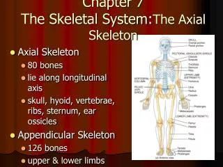
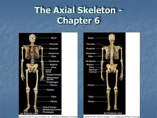
CHAPTER 7 THE SKELETON
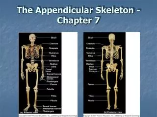
2Axial Skeleton: skull, vertebral column, thoracic cage Fig. 7.1 pg. 199
GO TO SLIDE 31 4 I. Skull: 2 parts A. Cranium: Encloses and protects the brain - 8 bones B. Facial bones: 14 bones Cranium A. Frontal – Forehead (brain) Anterior part of cranium forming the superior eye orbits then horizontal to form roof of eye orbit/floor of anterior cranial cavity Figs. 7.4 & 7. 5,Pgs. 202,203
9 B. Parietal bones (2) bilateral posterior, lateral 2/3 of cranium (brain) fig. 7.5 a C. Occipital: (brain) posterior/inferior part of the cranium fig. 7.4b; pg 202 & 7.6a; pg. 205 D. Temporal: (2) bilateral, inferior to parietals 1. houses: external auditory meatus (ear canal), middle & internal ear 2. Zygomatic process: posterior part of zygomatic arch Figures 7.5; pg 203 & fig. 7.8; pg. 207
12 Sutures: major, “immoveable” serrated joints between previous cranial bones (we’ll get back to remaining 2 skull bones latter) Sagittal suture: lies in the mid-sagittal plane & separates the left & right parietals Fig. 7.4b; pg. 202
14 B. Coronal suture: lies in coronal plane articulating both anterior parietals with the frontal bone fig. 7.5 a pg. 203 C. Lambdoid suture: posterior skull articulating both posterior parietals with the occipital bone . fig. 7.5 a pg. 203 D. Squamous suture: articulates inferior border of parietals w/ superior border of the temporal bones . Figure 7.5 a Cranial bones & sutures: prenatal & newborns Cranial bones cartilaginous [fontanelles] & sutures flexible - child birth
16 Last two cranial bones – cranial floor E. Sphenoid – middle part of cranial floor 1.Weird, bat shaped bone that articulates w/ all the other 7 cranial bones 2. Superior surface contains sella turcica (turk’s saddle) hold the pituitary gland (hypophysis) 3. Forms part of cranial floor 4. Part of external skull, anterior to temporal bone (greater wings) next slde Fig. 7.6a; pg 205/ Fig. 7.7a; pg. 206
19 F. Ethmoid bone: another bizarre shaped bone. 1. Location: midline in anterior part of cranial floor medial to orbits Figure 7.4a; page 202 2. Includes the cribiform plate a. part of the roof of the nasal cavity b. foramina (holes) that carry olfactory (smell) to brain
21 Facial Bones • Mandible – lower jaw bone 1. Coronoid (l) (crown shaped)“process” fig. 7.11 a pg. 210 a. Insertion of temporalis muscle – closes jaw 2. Mandibular condyle: articulates with mandibular fossa of temporal bone – forms tempromandibular joint - TMJ a. TMJ syndrome –symptoms from joint dyfunction next slide fig. 7.5a pg. 203
24 B. Maxillary bones: Upper jaw – 2, fused medially • “keystone” articulate w/ all other facial bones 2. part of floor of orbit (eye) 3. most of hard palate (roof of mouth) 4. floor of nasal cavity C. Zygomatic bone: “Cheek bone” zygoma (L)” – lateral wall & floor of orbit (eye socket) 1. Forms zygomatic arch w/ zygomatic process of temporal bone Fig. 7.5 & 7.6a&b pgs. 203 & 205
28 D. Nasal bones: Thin bones fused medially forming the bridge of the nose. a. Everything anterior to bridge is hyaline cartilage. E. Sinuses: 1. Hollow, cavities lined with mucous membranes – can become inflamed - allergies 2. Connect with nasal cavities 3. found in – frontal, sphenoid, ethmoid, maxillary bones
31 Hyoid bone: • Anchor for the tongue • Horseshoe shaped • Anchored to styloid processes by thin ligaments
33 Vertebral Column/Spinal Column Spine24 Vertebrae, Sacrum & Coccyx . Fig. 7.16; page 217 Three regions 1. Cervical: 7 vertebrae ; C1-C7 2. Thoracic: 12 vertebrae; T1-T12 3. Lumbar: 5 Vertebrae; L1- L5 4. Sacrum: 5 fused segments 5. Coccyx: 4 fused segments 3 Spinal Curves: 60 degrees 1. Cervical: forward curve & called a lordosis 2. Thoracic curve: reverse curve - kyphosis 3. Lumbar curve: forward curve - lordosis
35 Fetus and newborns have one, thoracic (kyphotic) curve – 2 other curves develop as child develops and becomes active Perfect Posture (biomechanical) Digress Vertebrae Structure 2 parts I. Body - large round thick bone disc a. Weight is transmitted from body to body c. Separated by intervertebral disc
Fig. 7.17 a; pg 218 Facet, neural/posterior arch 9th Ed 9th Ed 7.18 pg 219
37 Fig. 7.17 b; pg 218 Skip to 38 redundant DISC STRUCTURE, BULGE
39 II. Posterior (neural) arch – posterior to body A. Pedicles: project posteriorly from the body forming the lamina which meet medially forming the vertebral foramen B. Transverse processes: extend laterally for muscle attachment w/ leverage C. Spinous process: extends posteriorly for muscle attachment w/ leverage 1. Bumps down ones back
40 Fig. 7.18; page 219 9th Ed 7.19 pg. 220
E. Articular processes: 1. Have a smooth articular face called facet 2. Each vertebra has a superior articular process and an inferior articular process 3. The inferior facet of a vertebrae will articulate (meet) with the superior facet of the vertebra just inferior to it. 4. Facet joint: Where the facets articulate (meet) DIAGRAM 41 D. Inferior notch: notch for spinal nerve forming the intervertebral foramen, back to back to 36
43 lII. Vertebral Regions A. Cervical vertebrae Fig. 7.19 a&b; pg. 220 1. C1 – Atlas – holds up the head – Superior facets articulate w/ occipital condyles of skull a. No body or spinous process anterior/posterior tubercles 2. C2 – Axis 7.19 c; pg. 220 a. Has odontoid/dens post that acts as an axle for atlas to rotate on b. Majority of cervical rotation between C1 &C2
47 3. All cervical vertebrae, C1-C7, have transverse foramens for vertebral arteries . Table 7.2; pg. 222 4. Cervical vertebral bodies are small B. Thoracic Vertebrae T1-T12 . Table 7.2; pg. 222 1. Start out looking like cervicals T1 and become more like lumbars at T12 2. Facets lie in coronal plane – lateral flexion only 3. Have extra superior demifacets & inferior costal (rib) facets that articulate w/ ribs
50 C. Lumbar Vertebrae L1- L5 . 1. Largest, strongest vertebrae . 2. Facets lie in sagital plane forwards & . backwards only D. Sacrum S1 – S5 Solid fused mass . 1. Functional joint between L5 & S1 E. Coccyx - tail bone . 1. 4 fused segments . 2. Attached to sacrum by ligaments and . Can be sprained – coccydinia . 3. Vestigial tail – 1 of 100,000