Understanding Lipid Metabolism: Key Processes, Roles, and Transport Mechanisms
Lipid metabolism plays a crucial role in energy provision and cellular structure. Fatty acids, essential for heart and muscle energy, can be obtained from diet or synthesized. Triacylglycerides are efficient energy storage due to their high content, while bile salts facilitate their digestion through emulsification. Lipid digestion involves various enzymes and leads to the absorption of products via mixed micelles. The recycling of bile salts via enterohepatic circulation and the formation of chylomicrons ensure efficient transport of lipids in the bloodstream, preventing aggregation issues.

Understanding Lipid Metabolism: Key Processes, Roles, and Transport Mechanisms
E N D
Presentation Transcript
…Úrsula on the other hand, held a bad memory of that visit, for she had entered the room just as Melquíades had carelessly broken a flask of bichloride of mercury. “It’s the smell of the devil,” she said. “Not at all,” Melquíades corrected her. “It has been proven that the devil has sulphuric properties and this is just a little corrosive sublimate.” … Gabriel GarcíaMárquez, One Hundred Years of Solitude.
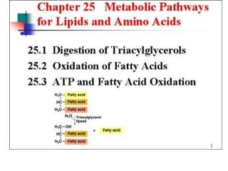
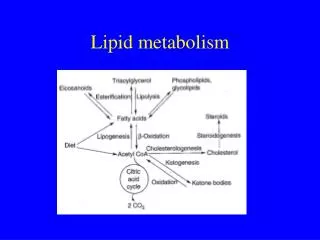
LIPID METABOLISM • Lipids serve as a source of energy and for the synthesis of membranes and biologically active molecules • Fatty acids are the preferred source of energy for the heart, the liver and for the skeletal muscles (during rest and prolonged activity) • Fatty acids can be obtained from the diet or they can be synthesized from excess carbohydrates and proteins (mainly in the liver and the adipose tissue) and stored in the adipose tissue • advantages to having triacylglycerides as storage lipids: • they have high energy content because they are highly reduced • they form aggregates because of their non-polar nature; osmolality is not raised and there is no solvation • stability of the C-C bonds
human milk, on the other hand, is rich in long-chain fatty acids which are essential for the development of the brain • some amount of lipases is also released along with the milk • The insolubility of lipids in water poses a problem for their digestion • lipid-digesting enzymes are water-soluble • they can access only the outer surface of the lipid aggregates • this problem is solved by bile salts which emulsify the lipids and increase the surface area that is exposed to the enzymes • Bile salts are amphipathic moleculessynthesized from cholesterol by the liver and stored in the gall bladder
The digestion and transport of lipids • Although the main lipids in the diet are triacylglycerides, small amounts of phospholipids, cholesterol and cholesterol esters are present • Most of the digestion of lipids in the intestine and produces free fatty acids, 2-monoacylglycerols, cholesterol and lysophospholipids (phospholipids with a fatty acid removed from carbon 2) • Lingual and gastric lipases are relatively active in infants and children • the enzymes cleave short and medium-chain ( < 12 carbons) fatty acids from triacyglycerides • short and medium-chain fatty acids are abundant in cow milk
Secretion of bile salts and cholesterol into the bile is the only way of excreting cholesterol • Bile salts enclose pieces of triacylglyceride • Colipase binds with TAG and lipase; removes bile salts and the lid that was covering the active site of lipase • Lipase breaks triacyglycerides down into two free fatty acids and 2-monoacyglycerol • Cholesterol esterase produces free cholesterol and a fatty acid molecule • Phospholipase A2 gives a lysophospholipid and a fatty acid • lysophospholipids are powerful detergents • assist in the emulsification process • Some amount of lecithin (phosphatidylcholine) is secreted in the bile • Bee and cobra venoms contain phospholipase A2
Short and medium-fatty acids are absorbed directly into the intestinal epithelial cells • Once in the cells, they are bound with intestinal fatty acid binding protein (I-FABP)
From the intestine they pass to the portal veins and they travel to the liver bound with albumin • the function of I-FABP and albumin is to increase solubility and prevent the damage to cell membranes that could be brought by the detergent properties of free fatty acids • The absorption of the remaining products of lipid digestion is through the formation of mixed micelles that are made up of amphipathic molecules: bile salts, lysophospholipids, long-chain fatty acids, 2- monoacyglycerol , cholesterol and fat-soluble vitamins • Except for the bile salts, the rest are absorbed by the duodenal and jejunal epithelial cells • in the epitheliumtriacylglycerides, phospholipids and some cholesterol ester are resynthesized • > 95 % of the bile salts reabsorbed in the ileum and returned to the liver (enterohepatic circulation) to be recycled; the rest is excreted
Absorption of the products of lipid digestion
Enterohepatic circulation of bile salts
Steatorrhea or the excretion of undigested lipids could result from the deficiency of pancreatic lipase or bile salts • the digestion and absorption of fat-soluble vitamins is also impaired • If the resynthesized TAG were to enter the blood directly, they would have formed aggregates and blood flow would have been blocked • This problem is solved through the synthesis of chylomicrons – one type of lipoproteins • The idea behind lipoproteins is to transport hydrophobic lipids by sequestering them in the core of the particle and covering them with a hydrophilic layer made up of proteins • The core of chylomicrons consists of TAG, cholesterol esters and vitamins; the surface is made of proteins and the polar groups of cholesterol and phospholipids
The resynthesis of TAG • The main apoproteinof chylomicrons is Apo B-48 and is produced by the rough endoplasmic reticulum • TAG resynthesized by the smooth ER; TAG and Apo B-48 are packed into chylomicrons in the Golgi complex • Other proteins in chylomicrons include Apo-CII and Apo-E • Of the lipoproteins, chylomicrons are the biggest in the size and the lowest in density (smallest amount of proteins)
Nascent chylomicrons(containing only Apo B-48) are exocytosed into the lymph and then join the blood stream • While in the circulation, they receive the other apoproteins from high density lipoprotein (HDL) and become mature chylomicrons • a fat-rich meal gives the blood a milky appearance • Chylomicrons are destined mainly for the adipose, muscles (especially cardiac muscles) and lactating mammary glands • These tissues produce lipoprotein lipase (LPL) • LPL is found associated with the proteoglycans in the basement membranes of the endothelial cells of the capillaries serving these tissues • The LPL isozyme from the adipose has a higher Km and is most active after meals when there is much TAG in the blood • insulin stimulates the synthesis of adipose LPL
LPL is activated by Apo CII and removes fatty acids from the TAG of the chylomicrons • The free fatty acids travel bound with albumin until they are absorbed • The fatty acids are used as energy sources in the muscles • In the adipose they are reesterified and stored while in the mammary glands they are incorporated into milk fat • The glycerol that was produced through the action of LPL travels to the liver and could be used for TAG synthesis in the fed-state • When most of the TAG has been removed, chylomicrons shrink and become chylomicron remnants • Chylomicron remnants are endocytosed by the liver (through recognition of Apo E) and their contents are degraded in the lysosomes
Mobilization of triacylglycerides from the adipose • The adipose stores mainly TAG and steroid hormone – synthesizing tissues like the adrenal cortex, ovaries and testes store cholesterol esters • The lipids are stored in the form of lipid droplets: a core of TAG or cholesterol esters surrounded by a single layer of phospholipids and perilipinproteins • Perilipins prevent the untimely mobilization of lipids • The release of glucagon in the fasting state or epinephrine in the fight or flight response increases the level of cAMP– a secondary messenger – in the adipose • cAMP activates protein kinase A which in turn phosphorylatesperilipin • The phosphorylatedperilipin allows hormone-sensitive lipase (HSL) to move to the surface of the lipid droplet
HSL also is phosphorylated by protein kinase A; but the main factor for the massive increase in mobilization is the phosphorylation of perilipin • HSL breaks TAG down into fatty acids and glycerol • The glycerol travels to the liver and is used for gluconeoge. • Some of the fatty acids travel-bound with albumin-to the cardiac and skeletal muscles and the renal cortex; provide energy • About 75 % of the fatty acids released by lipolysis are reesterified • This is known as the triacylglycerol cycle • the fatty acids are reesterified while they are still in the adipose or after they have travelled to the liver • The cycle operates even during starvation
In the adipose tissue, dihydroxyacetone from glycolysis is changed to glycerol-3-phosphate for TAG synthesis; the liver can make use of glycerol • But glucagon and epinephrine inhibit glycolysis and DHAP would not be available in the adipose for TAG synthesis • What is the source of the DHAP used for the synthesis of TAG in the adipose during starvation? • glyceroneogenesisis a shortened version of gluconeogenesis thatproduces DHAP from gluconeogenic substrates like alanine, aspartate and malate • the adipose has two gluconeogenic enzymes – pyruvate carboxylaseand PEP carboxykinase – even though it does not produce glucose
Glyceroneogenesis supports TAG synthesis in the liver during starvation • Natural and artificial glucocorticoids induce the synthesis of the liver isozyme of PEP carboxykinase while inhibiting the adipose isozyme • little reesterification in the adipose; more fatty acids will be released to the blood stream • but esterification in the liver will be more active and reestablish the 75 % • Excess levels of fatty acids in the blood have been implicated in the development of insulin resistance • thiazolidinedionesare drugs that induce the synthesis of the adipose isozyme of PEP carboxykinase • used in the treatment of type 2 diabetes mellitus
The triacylglycerol cycle Thiazolidinediones
FATTY ACID CATABOLISM/β-OXIDATION • Fatty acids released from the adipose have their origins in the diet or synthesis in the liver • The most common dietary fatty acids are palmitate (16:0), stearate (18:0), oleate (18:1) and linoleate (18:2) • Animal fat contains mostly saturated and monounsaturated fatty acids while vegetable oils contain linoleate and longer polyunsaturated fatty acids • Fatty acids enter cells both by diffusion and sodium- dependent transport • As shown by Lehninger and Kennedy, the oxidation of fatty acids takes place in the mitochondria • Short and medium-chain fatty acids can freely enter into and be activated in the mitochondria
long chain fatty acids, on the other hand, are first activated in a reaction with CoA; form a high energy thioester • the acyl-CoAsynthetaseis present in the ER and the outer membranes of the mitochondria and the peroxisomes . It shows little or no activity with short, medium and very long chain (>22C) fatty acids
The fatty acyl-CoA can be used either in the synthesis of TAG and membrane lipids or transported into the mitochondria and used for energy production • Transport by the carnitine shuttle • Carnitine reacts with fatty acyl-CoA and gives acyl- carnitine; commits the fatty acids to oxidation • the enzyme responsible is carnitine acyltransferase I (CAT I) which is located on the outer mitochondrial membrane • Acylcarnitine crosses the inner membrane through a translocase • In the mitochondria CAT II changes acylcarnitine to fatty acyl-CoA • Carnitine returns to the cytosol through the translocase
As the name indicates, β-oxidation is a set of reactions aimed at forming a carbonyl group on the β-carbon • The presence of two carbonyl groups weakens the otherwise stable Cα-Cβ bond and acetyl-CoA groups are removed successively • One cycle ofβ-oxidation consists of four reactions • The oxidation of the Cα-Cβbond • Catalyzed by acyl-CoAdehydrogenases-a family of three isozymes specific for short, medium and long-chain FA • Contain non-covalently (but tightly) attached FAD • FADH2 transfers the electrons to electron transfer flavoprotein (ETF) • ETF is oxidized by ETF:UQ oxidoreductase and the electrons are delivered to coenzyme Q
Addition of water across the double bond • Trans-Δ2-enoyl-CoA is acted upon by enoyl-CoAhydratase • Oxidation of the β–hydroxyl group • β–hydroxyacyl-CoA is oxidized by L-hydroxyacyl-CoA dehydrogenase; NAD is the electron carrier
cleavage of the Cα-Cβ bond • β–ketoacyl-CoAcleaved by β–ketoacylthiolase • An acetyl-CoA molecule and an acyl-CoA two carbons shorter are produced • Steps 2-4 of the β-oxidation of long chain fatty acids are carried out by a multienzyme complex known as trifunctional protein (TFP) • individual enzymes for short and medium chain FA
A cycle of β-oxidation releases one molecule each of FADH2,NADH and acetyl-CoA • the four steps are repeated n/2 - 1 times ( n = number of carbons) until the final acetyl-CoA is released • n/2 acetyl-CoA and n/2 - 1 each of FADH2 and NADH are produced by the complete oxidation of a fatty acid with an even number of carbons • taking the oxidation of palmitate (16:0): • Palmitoyl-CoA+ 7 CoA+ 7 FAD+ 7 NAD++7 H2O→ 8 acetyl-CoA+ 7 FADH2+7 NADH+ 7H+ • 8 acetyl-CoA ≈ 8o ATP, 7 FADH2 ≈ 10.5 ATP and 7 NADH ≈ 17.5 ATP ⇒ total = 108 ATP • the equivalent of two ATP is spent on activation of FA • ⇒ net production of 106 ATP • The oxidation of fatty acids releases a large amount of metabolic water
the β-oxidation of odd-chain fatty acids • Odd-chain FA are common in plants and marine organisms • β-oxidation proceeds in the normal way but the last product to be released is not acetyl-CoA but propionyl-CoA • The propionyl-CoA is first changed to D-methylmalonyl-CoA through the action of propionyl-CoAcarboxylase • biotin is the coenzyme • D-methylmalonyl-CoA is epimerized • L-methylmalonyl-CoA undergoes intramolecular rearrangementto give succinyl-CoA • this is one of the two reactions in humans where vitamin B 12 is involved • Succinyl-CoA can be used for energy production or the synthesis of glucose
the β-oxidation of unsaturated fatty acids • Double bonds naturally occurring in fatty acids are in the cisform • Enoyl-CoAhydratase cannot act on cisdouble bonds • In order for β-oxidation to proceed, either the cisform has to be changed to trans or the double bond should be reduced • Two additional enzymes are needed – an isomerase and a reductase • For monounsaturated fatty acids, the enoyl-CoAisomerase suffices • Polyunsaturated fatty acids need the participation of both enzymes • NADPH is used as the reductant
This slide: the oxidation of a monounsaturated fatty acid – oleate Next slide: the oxidation of a polyunsaturated fatty acid–linoleate
the regulation of β-oxidation • The concentration of mitochondrial CoA may be limiting to the formation of fatty acyl-CoA • The formation of malonyl-CoA is the first step during the synthesis of fatty acids • Malonyl-CoA inhibits CAT I making sure that fatty acids do not get degraded at the same time they are synthesized • High levels of FADH2 and NADH respectively inhibit acyl-CoA dehydrogenase and β-hydroxyacyl-CoA dehydrogenase; high levels of acetyl-CoA inhibit thiolase
Disorders of fatty acid oxidation • Inherited defects in the carnitine-assisted transport or the enzymes of β-oxidation are manifested as serious heart disease • CAT I/II, carnitine translocase, acyl-CoAdehydrogenases, … may be defective • Carnitine obtained from the diet or synthesized in the body • carnitine deficiency is unlikely to be seen in adults • carnitine related enzymes and transporters are more commonly deficient leading to the accumulation of long chain acyl/ acyl carnitine • weak muscles; should avoid prolonged exercise and fasting • Acyl-CoA dehydrogenase deficiency results in the excretion of *derivatives of fatty acids in the urine and *non-ketotic hypoglycemia
Alternate routes of fatty acid oxidation • Minor, yet important pathways of fatty acid oxidation • peroxisomaloxidation • Human diet usually contains VLCFA and branched-chain fatty acids from chlorophyll degradation • VLCFA are also produced in high amounts in the nervous system and incorporated into the myelin sheath • VLCFA undergo β-oxidation but the first step is unique in that it transfers electrons from acyl-CoA to FAD and then to O2 - production of H2O2 Acyl-CoAoxidase
The three remaining reactions of β-oxidation proceed in the normal way • Acetyl groups move out of the peroxisome by themselves or carried by carnitine • When the acyl-CoA is shortened to about 8 carbons, it is transported out of the peroxisomes (with carnitine) and its oxidation is completed in the mitochondria
Another type of oxidation takes place in the peroxisomes – • α-oxidation of branched-chain fatty acids • Mainly on phytanic acid and pristanic acid • phytanic acid is first changed to pristanic acid by hydroxylation of the α carbon followed by decarboxylation • The shortening by one carbon puts methyl groups on the α rather than the βcarbon –normal β oxidation continues • Acetyl-CoA and propionyl-CoA are alternatively removed from the acyl-CoA • When a middle-chain acyl-CoA is reached, it is transported to the mitochondria • Refsum’s disease - deficiency of phytanic acid αhydroxylase • accumulation of phytanic acid in the body • leads to serious neurological problems
Zellweger syndrome: the absence of functional peroxisomes ; X-linked adrenoleukodystrophy (XALD): lack of tansporters for VLCFA into the peroxisomes • accumulation of VLCFA in the body, especially C26:0 and C26:1 • Affect mainly the liver and the brain; fatal in the very early years of life • ω-oxidation • In the endoplasmic reticulum, the end farthest from the carboxyl group is oxidized • first, the methyl group is changed to hydroxyl by cytochrome p 450 enzymes • Then comes dehydrogenation which yields a dicarboxylic acid
Either end can be attached to CoA and undergo β oxidation • β oxidation produces smaller dicarboxylic acids such as adipicand succinic acids • Dicarboxylic acids are highly water-soluble and can be removed through the urine • ω-oxidation is normally involved in the metabolism of xenobiotics that have structures similar with fatty acids • ω-oxidation becomes more prominent when β oxidation is defective and the need arises to excrete the unmetabolized fatty acids
Metabolism of ketone bodies • Most of the acetyl-CoA produced from hepatic fatty acid oxidation enters the TCA cycle • The remaining acetyl-CoA molecules undergo ketogenesis – the production of the ketone bodies: acetone, acetoacetateand β-hydroxybutyrate • The ketone bodies are released from the liver and provide energy for the heart, skeletal muscles, renal cortex and brain • changed back into acetyl-CoA and join the TCA cycle • Ketogenesis occurs in the mitochondrial matrix • two acetyl-CoA condense to give acetoacetyl-CoA • another acetyl-CoA is added to acetoacetyl-CoA to give β-hydroxyl-β –methyl –glutaryl –CoA (HMG-CoA) • HMG-CoA is changed to acetoacetate and acetyl-CoA • Acetoacetate is reduced to β-hydroxybutyrate or decarboxylated to acetone
The thiolasethat catalyzes the first reaction of ketogenesis is the same enzyme that is involved in the last reaction of βoxidation • Ketogenesisand *cholesterol synthesis share the reactions up to the formation of HMG-CoA • Acetoacetate and β-hydroxybutyrate are exported to other tissues through the blood • ketone bodies are easily transportable forms of fatty acids • the conversion of acetoacetate and β-hydroxybutyrate back to acetyl-CoA takes place in the mitochondria • two reactions are retained from ketogenesis and one new reaction is introduced – the activation of acetoacetate by the transfer of CoA from succinyl-CoA • β-ketoacyltransferaseis absent in the liver • Most of the acetone is exhaled
2 acetyl CoA = 20 ATP • changing succinylCoA to succinate • without the production of GTP • ⇒ subtract 1 ATP for the activation • 19 ATP • starting fromβ-hydroxybutyrate • ⇒ 21.5 ATP (additional energy from • NADH)
The degradation of *ketogenic amino acids provides acetoacetyl-CoAand acetyl-CoA • The abundance of fatty acids in the blood during the fasting state increases the level of ketone body synthesis • Oxaloacetate is diverted to gluconeogenesis • More acetyl-CoA enters ketogenesis because of the small levels of oxaloacetate to condense with (for TCA) • In type I diabetes, the inhibitory effect of insulin on lipolysis is relieved • fatty acids released by lipolysis in turn produce excess amounts of acetyl-CoA • even though glucose is abundant in the blood gluconeogenesis continues unabated – oxaloacetate is consumed for glucose production