1.3 Membrane Structure
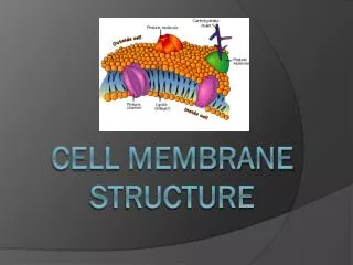
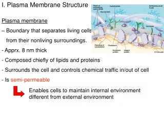
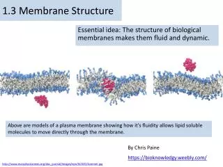
1.3 Membrane Structure. Essential idea: The structure of biological membranes makes them fluid and dynamic. Above are models of a plasma membrane showing how it's fluidity allows lipid soluble molecules to move directly through the membrane. By Chris Paine https :// bioknowledgy.weebly.com /.

1.3 Membrane Structure
E N D
Presentation Transcript
1.3 Membrane Structure Essential idea: The structure of biological membranes makes them fluid and dynamic. Above are models of a plasma membrane showing how it's fluidity allows lipid soluble molecules to move directly through the membrane. By Chris Paine https://bioknowledgy.weebly.com/ http://www.europhysicsnews.org/doc_journal/images/epn/hl/435/Sommer.jpg
1.3.U1 Phospholipids form bilayers in water due to the amphipathic properties of phospholipid molecules. What happens when you put a drop of oil in water? http://www.flickr.com/photos/zorin-denu/5385963280/
1.3.U1 Phospholipids form bilayers in water due to the amphipathic properties of phospholipid molecules. The Oil droplet stays together and makes a perfect circular shape. • The oil molecules are • Hydrophobic • Oil Molecules are non-polar and water • molecules are polar. • See 3.1.5 http://www.flickr.com/photos/zorin-denu/5385963280/
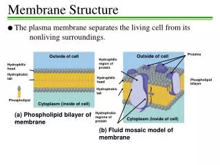
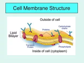
1.3.U1 Phospholipids form bilayers in water due to the amphipathic properties of phospholipid molecules. • Phospholipid moleculeshave a polar (charged) phosphate head and long non-polar lipid tails • The head is hydrophillic (attracted to water) • The tails are hydrophobic (repelled by water) When drawing a diagram of a phospholipid this is a good example which shows all the key features http://www.ib.bioninja.com.au/_Media/phospholipid_bilayer_med.jpeg http://upload.wikimedia.org/wikipedia/commons/3/39/Phospholipid_TvanBrussel.jpg
1.3.U1 Phospholipids form bilayers in water due to the amphipathic properties of phospholipid molecules. When put into water, an emergent property is that phospholipids will self-organise to keep their heads ‘wet’ and their tails ‘dry’ micelle liposome http://commons.wikimedia.org/wiki/File:Micelle_scheme-en.svg http://commons.wikimedia.org/wiki/File:Liposome_scheme-en.svg
1.3.U1 Phospholipids form bilayers in water due to the amphipathic properties of phospholipid molecules. In this 3D representation you can see that a phospholipid bilayer is one way that the tails can be removed from the water. Phospholipid molecules can flow past each other laterally but can’t move vertically http://commons.wikimedia.org/wiki/File:Phospholipids_aqueous_solution_structures.svg
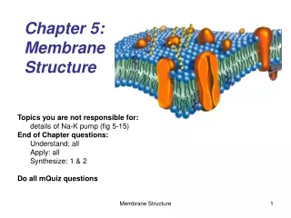
1.3.U1 Phospholipids form bilayers in water due to the amphipathic properties of phospholipid molecules. But wait! there’s more! The plasma membrane is not just made of phospholipids http://commons.wikimedia.org/wiki/File:Cell_membrane_detailed_diagram_en.svg?uselang=en-gb
1.3.U2 Membrane proteins are diverse in terms of structure, position in the membrane and function. Proteins: Integral proteins are permanently embedded, many go all the way through and are polytopic (poly = many, topic = surface), integral proteins penetrating just one surface are monotopic. Peripheral proteins usually have a temporary association with the membrane, they can be monotopic or attach to the surface http://commons.wikimedia.org/wiki/File:Cell_membrane_detailed_diagram_en.svg?uselang=en-gb
1.3.U2 Membrane proteins are diverse in terms of structure, position in the membrane and function. Glycoproteins: Are proteins with an oligosaccaride (oligo = few, saccharide = sugar) chain attached. They are important for cell recognition by the immune system and as hormone receptors http://commons.wikimedia.org/wiki/File:Cell_membrane_detailed_diagram_en.svg?uselang=en-gb
1.3.U2 Membrane proteins are diverse in terms of structure, position in the membrane and function. Transport: Protein channels (facilitated) and protein pumps (active) Receptors: Peptide-based hormones (insulin, glucagon, etc.) Anchorage: Cytoskeleton attachments and extracellular matrix Cell recognition: MHC proteins and antigens Intercellular joinings: Tight junctions and plasmodesmata Enzymatic activity: Metabolic pathways (e.g. electron transport chain) http://www.ib.bioninja.com.au/standard-level/topic-2-cells/24-membranes.html
1.3.U3 Cholesterol is a component of animal cell membranes. Cholesterol: (It’s not all bad!) It makes the phospholipids pack more tightly and regulates the fluidity and flexibility of the membrane. Bad analogy: imagine a room full of people wearing fluffy jumpers (sweaters). It is crowded but they can slip past each other easily enough. Now sprinkle the crowd with people wearing Velcro™ suits…
1.3.U3 Cholesterol is a component of animal cell membranes. Cholesterol • Hydroxyl group makes the head polar and hydrophilic - attracted to the phosphate heads on the periphery of the membrane. Carbon rings – it’s not classed as a fat or an oil, cholesterol is a steroid Non-polar (hydrophobic) tail –attracted to the hydrophobic tails of phospholipids in the centre of the membrane http://www.uic.edu/classes/bios/bios100/lectf03am/cholesterol.jpg http://www.cholesterol-and-health.com/images/Cholesterol_Structure.jpg
1.3.A1 Cholesterol in mammalian membranes reduces membrane fluidity and permeability to some solutes. Membrane fluidity • The hydrophobic hydrocarbon tails usually behave as a liquid. Hydrophilic phosphate heads act more like a solid. • Though it is difficult to determine whether the membrane is truly either a solid or liquid it can definitely be said to be fluid. • It is important to regulate the degree of fluidity: • Membranes need to be fluid enough that the cell can move • Membranes need to be fluid enough that the required substances can move across the membrane • If too fluid however the membrane could not effectively restrict the movement of substances across itself http://www.nature.com/scitable/content/ne0000/ne0000/ne0000/ne0000/14668965/U2.cp5.3_membrane_f2.jpg
1.3.A1 Cholesterol in mammalian membranes reduces membrane fluidity and permeability to some solutes. Cholesterol’s role in membrane fluidity • The presence of cholesterol in the membrane restricts the movement of phospholipids and other molecules– this reduces membrane fluidity. 1. • The presence of cholesterol disrupts the regular packing of the of the hydrocarbon tails of phospholipid molecules - this is increases the flexibility as it prevents the tails from crystallising and hence behaving like a solid. 2. Cholesterol also reduces the permeability to hydrophilic/water soluble molecules and ions such as sodium and hydrogen. 3. http://www.nature.com/scitable/content/ne0000/ne0000/ne0000/ne0000/14668965/U2.cp5.3_membrane_f2.jpg
1.3.S1 Drawing of the fluid mosaic model. Use the tutorials to learn and review membrane structure http://www.bio.davidson.edu/people/macampbell/111/memb-swf/membranes.swf http://www.phschool.com/science/biology_place/biocoach/biomembrane1/regions.html https://www.wisc-online.com//LearningContent/ap1101/index.html
1.3.S1 Drawing of the fluid mosaic model. http://www.youtube.com/watch?v=w9VBHGNoFrY
1.3.S1 Drawing of the fluid mosaic model. Reminder of features that make good diagrams: • Good use of space • Clear strong lines • Label lines are straight • Labels clearly written • (Scale bar if appropriate) • Lines touch the labeled structure • No unnecessary shading or colouring http://www.ib.bioninja.com.au/_Media/phospholipid_bilayer_med.jpeg
1.3.S3 Analysis of the falsification of the Davson-Danielli model that led to the Singer-Nicolson model. Our current model of the cell membrane is called the Singer-Nicholson fluid mosaic model • Key features: • Phospholipid molecules form a bilayer - phospholipids are fluid and move laterally • Peripheral proteins are bound to either the inner or outer surface of the membrane • Integral proteins - permeate the surface of the membrane • The membrane is a fluid mosaic of phospholipids and proteins • Proteins can move laterally along membrane http://commons.wikimedia.org/wiki/File:Cell_membrane_detailed_diagram_en.svg?uselang=en-gb
1.3.S3 Analysis of the falsification of the Davson-Danielli model that led to the Singer-Nicolson model. • Our current model of the cell membrane is called the Singer-Nicholson fluid mosaic model • There is strong evidence for this model: Biochemical techniques • Membrane proteins were found to be very varied in size and globular in shape • Such proteins would be unable to form continuous layers on the periphery of the membrane. • The membrane proteins had hydrophobic regions and therefore would embed in the membrane not layer the outside http://commons.wikimedia.org/wiki/File:Cell_membrane_detailed_diagram_en.svg?uselang=en-gb
1.3.S3 Analysis of the falsification of the Davson-Danielli model that led to the Singer-Nicolson model. • Our current model of the cell membrane is called the Singer-Nicholson fluid mosaic model • There is strong evidence for this model: Fluorescent antibody tagging • red or green fluorescent markers attached to antibodies which would bind to membrane proteins • The membrane proteins of some cells were tagged with red markers and other cells with green markers. • Within 40 minutes the red and green markers were mixed throughout the membrane of the fused cell. • This showed that membrane proteins are free to move within the membrane rather than being fixed in a peripheral layer. • The cells were fused together. http://commons.wikimedia.org/wiki/File:Cell_membrane_detailed_diagram_en.svg?uselang=en-gb
1.3.S3 Analysis of the falsification of the Davson-Danielli model that led to the Singer-Nicolson model. Our current model of the cell membrane is called the Singer-Nicholson fluid mosaic model This model was first proposed in by Singer-Nicolson in 1972 Before then Davson-Danielli model was widely accepted … http://commons.wikimedia.org/wiki/File:Cell_membrane_detailed_diagram_en.svg?uselang=en-gb
1.3.S2 Analysis of evidence from electron microscopy that led to the proposal of the Davson-Danielli model. • The evidence: In high magnification electron micrographs membranes appeared as two dark parallel lines with a lighter coloured region in between. • Proteins appear dark in electron micrographs and phospholipids appear light - possibly indicating proteins layers either side of a phospholipid core. Davson-Danielli Model Pore Proteins • The model: • A protein-lipid sandwich • Lipid bilayer composed of phospholipids (hydrophobic tails inside, hydrophilic heads outside) • Proteins coat outer surface • Proteins do not permeate the lipid bilayer Phospholipids This explains: Despite being very thin membranes are an effective barrier to the movement of certain substances. http://www.cytochemistry.net/cell-biology/EMview.jpg http://upload.wikimedia.org/wikipedia/commons/6/69/Davson_danielli_miguelferig.jpg
1.3.S3 Analysis of the falsification of the Davson-Danielli model that led to the Singer-Nicolson model. Falsification of the Davson-Daniellimodel – freeze fracturing • Interpreting the image: • The fracture occurs along lines of weakness, including the centre of membranes. • The fracture reveals an irregular rough surface inside the phospholipid bilayer • The globular structures were interpreted as trans-membrane proteins. • This technique involves rapid freezing of cells and then fracturing them. Conclusion: This is contrary to the Davson-Danielli model which only involves proteins coating the surface of the membrane. A new model is needed to explain the presence of as trans-membrane proteins. http://www.cytochemistry.net/cell-biology/ffimage.jpg
Bibliography / Acknowledgments Jason de Nys