The Liver & Gallbladder
The Liver & Gallbladder. The liver has been shown to have m ore than 500 vital functions We will review only a few of these. PRODUCES BILE Elimination of toxins F at emulsifier H elps alkalinize SI with HCO 3 Aids in vit K absorption from gut CLOTTING

The Liver & Gallbladder
E N D
Presentation Transcript
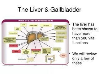
The Liver & Gallbladder • The liver has been shown to have more than 500 vital functions • We will review only a few of these
PRODUCES BILE Elimination of toxins Fat emulsifier Helps alkalinize SI with HCO3 Aids in vit K absorption from gut CLOTTING Produces clotting factors, prothrombin& fibrinogen (+vit K above) ENDOCRINE FUNCTIONS Secretes Insulin like growth factor (IGF-1) aka SomatomedinC Converts 60% of T4 >T3 IMMUNE FUNCTIONS Kupffer Cells ingest old RBCs, WBCs, viruses, and bacteria that enter though the small intestine Produces Complement NUTRIENT STORAGE & RELEASE Stores Glycogen, makes glucose Produces cholesterol Makes apoproteins for fats to travel around the body Stores vitamin ADEK Regulates amino acid levels in blood stores Fe (ferritin), Cu ACTIVATION OF VIT D Liver adds 1st OH to form hydroxy vitamin D3 Produces blood plasma proteins (albumin) that create the blood colloid osmotic pressure DETOXIFICATION Makes fat soluble drugs, hormones, waste products water soluble & excretes it through bile Converts poisonous ammonia from protein metabolism into urea Main Functions of the Liver
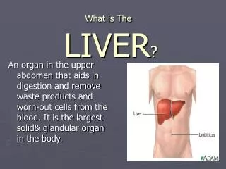
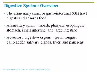
Anatomy • Largest organ in body (~3 lb) • located in the right upper quadrant, behind ribs, between the fifth intercostal space (below nipple) at the midclavicular line and the right costal margin. • It extends across the midline. • The falciform ligament divides the liver into right & left lobes & suspends the liver from the diaphragm
Hepatic circulation • 70% of the blood supply to the liver comes from the hepatic portal vein, whichcarries blood filled with absorbed nutrients from digestive organs to liver. • Oxygen-rich blood enters from the hepatic artery and mixes with the blood from the hepatic portal vein in the sinusoids • Blood exits the liver via the hepatic vein to the vena cava
Liver lobule • The liver is divided into hexagonal units filled with rows of hepatocytes called lobules. • At each corner of the hexagon, there is a portal triad which consists of a branch from (1) the hepatic artery, (2) the hepatic portal vein, and (3) a bile duct. • The blood from the hepatic artery and the hepatic portal vein mixes together in sinusoids and flows towards a central vein.
Liver Lobule 3D Blood is flowing up the central vein towards the vena cava. Bile is flowing down towards the gallbladder.
Vitamin A Storage & Stellate Cells • Stellate cells (Ito) lie between the sinusoids and the hepatocytes in the Space of Disse • Normally, they store Vitamin A in lipid droplets • In the presence of long term inflammation, they activate, transform into fibrogenic cells, leading to fibrosis of the liver
Kuppfer Cells • The liver has the largest amount of Resident macrophages in the body • phagocytize old RBCs • efficiently scavenge bacteria & substances that get into portal venous blood through breaks in the intestinal epithelium, thus preventing invasion of the systemic circulation • Responsible for liver damage in alcoholics
METABOLIC FUNCTIONS of the LIVER • Carbohydrate: • Maintains normal blood glucose levels • Glycogenolysis, Glycolysis, Gluconeogenesis (makes glucose from non-sugars eg amino acids, lactic acid, …) • Amino acids • Deamination: Removes NH2 from amino acids so they can be used as fuel • Converts toxic NH3 (ammonia) to less toxic urea • Fats • Synthesizes lipoproteins –VLDL, HDL • Synthesizes cholesterol
Insulin & Glucagon Glucose & glycogen
Glycogenesis vsGlycogenolysis • Glycogenesis:when blood glucose is high, pancreas secretes insulin, and liver converts excess blood glucose into glycogen (starch granules) • Glycogenolysis:if blood glucose is low, the pancreas secretes glucagon, and the liver will break down glycogen into glucose and release it into the blood. • Gluconeogenesis: In conditions of starvation (ie glycogen has been depleted), the liver can make glucose from other sources, like fats or proteins.
Insulin vs. Glucagon Insulin promotes • Glucose uptake • Glycogenesis: making glycogen • Lipogenesis: making fats Glucagon promotes • Glycogenolysis: breakdown of glycogen into glucose • Amino acid catabolism • Ketogenesis
Transamination, Ammonia, Urea Protein, amino acids
20 Amino Acids • There are 20 amino acids. 10 are essential - ie must get them from the diet. • An amino acid can only be used as a fuel source if the nitrogen, or amino group, NH3, is removed. NH3 is toxic to the brain so it must be excreted
Aminotransferases (ALT, AST) • Amino acids, like alanine, aspartate, glutamate, without the amino group, are called α-keto acids. Examples: pyruvate, oxaloacetate, α-ketoglutarate • Aminotransferase enzymes transfer amino groups between amino acids and keto acids. This is called transamination. • Aminotransferases usually use glutamate as the donor of an amino group, or its complementary keto acid, α-ketoglutarateas the acceptor of NH2
Keto acids enter the Krebs Cycle • Amino acids are transaminated to make keto acids. Each keto acid can enter the Krebs Cycle at its appropriate points. • this is reversible – krebs intermediates can also be used to make Aas • In this way, the liver makes it possible to use proteins as an energy source, when glucose is not available.
Alanine from muscles for Gluconeogenesis • During starvation(when glycogen stores are depleted in the liver), muscles will break down its proteins into AAs and form the AA, glutamate. • Alanine aminotransferase enzyme in muscles will transfer an amino group from glutamate, to turn pyruvate into alanine. • Alanine will now travel through the blood to liver, which can convert it into back into pyruvate using Alanine Aminotransferase (ALT). With new supply of pyruvate, the liver can make glucose (gluconeogenesis) to feed the brain.
Amino Acid Deamination • The liver takes 1 ammonium ion NH4 + from glutamate & a 2nd amino group from aspartate, and combines them to make urea, which has 2 amino groups. Urea can be excreted. • ammonia is toxic to the brain & must be converted to the less toxic urea
Urea Cycle FYI Glutamate donates one ammonium ion (which converts ornithine into citrulline). Aspartate donates the second ammonium ion (making citrulline into arginosuccinate)
Cholesterol • Cholesterol in the liver comes from • dietary sources (chylomicrons) • or can be made de novo by the liver. • Too much cholesterol entering hepatocytes from the LDLs will inhibit further synthesis of both cholesterol & LDL receptors • Cholesterol is removed from the liver by VLDLs
Some Cholesterol Uses • Cholesterol is the precursor for various substances in the body and needs to be delivered to many tissues for this function. Above, are some substances synthesized from cholesterol in the adrenal cortex.
Lipoproteins - Lipid Transport • Since cholesterol and other fats are insoluble in blood, they must be transported through the circulatory system as lipoproteins. • The liver makes lipoproteins,a combination of droplets of lipids + Apoproteinsin the outer shell: • VLDL - very low density lipoprotein (TG) • LDL - low density lipoprotein (cholesterol) • IDL - intermediate density lipoprotein • HDL - high density lipoprotein • Names reflect the amount of proteincontent. HDL has most.
Phospholipids • Most phospholipids are synthesized on the smooth endoplasmic reticulum of the hepatocytes • The most abundant phospholipid (50%) is phosphatidylcholine aka lecithin
Phospholipids • Phosphatidylcholine, along with bile salts, is needed to make the outer monolayer of micellesthat carry dietary emulsified fats. • PC is also critical to VLDL formation to transfer fat out of the liver & for other lipoprotein formation
Plasma proteins • 12 proteins comprise 96% of the plasma proteins. • Albumin, which is made by the liver, is, by far, the most abundant
FYI - Liver makes many Plasma Proteins Major plasma proteins: • Albumin, α-fetoprotein, (fetal albumin), Soluble plasma fibronectin, C-reactive protein, acute phase protein, various globulins Proteins of hemostasis and fibrinolysis • Coagulation: All coagulation cascade factors, except VIII (from endothelium); • Inhibitors of coagulation: α2-macroglobulin,α1-antitrypsin, Antithrombin III, Protein S, Protein C; • Fibrinolysis:(clot dissolution) plasminogen; Inhibitors of fibrinolysis: α2-antiplasmin; Immune Factors: Complement components C1-9, Complement component 3 (C3) Carrier Proteins & Binding Globulins: • Albumin - main carrier protein, carries hormones (including thyroid), fatty acids to the liver, unconjugated bilirubin, many drugs and Ca2+ • Sex hormone-binding globulin (testosterone, estradiol); Thyroxine-binding globulin (T4 and T3), Transferrin (ferric form Fe3+); Ceruloplasmin (Cu); Vitamin D binding protein Hormones: Insulin-like growth factor 1 (childhood growth) Thrombopoietin (produce platelets) Prohormones: Angiotensinogen (blood pressure) Apolipoproteins - all except Apo-B48 (produced by intestine)
Phase I & phase II Detoxification
Detoxification • The liver eliminates fat soluble toxins & hormones from the blood in 2 steps: phase I & phase II • Phase I produces less lipophillic substances but sometimes also dangerous free radicals and more toxic intermediaries • Phase II adds a substance to the toxin to make it water soluble and able to excrete in bile /feces & urine
Phase I: Cytochrome enzymes, CYP • The liver uses a super family of enzymes, called cytochromes or p450 usually located on the endoplasmic reticulum, to catalyze phase I reactions such as oxidation, reductions or hydolysis • Phase I modifies both exogenous (such as drugs, herbs, and pestcides) and endogenous toxins (such as hormones) into intermediate substances.
CYP Induction & Inhibition • One cytochrome isoform can ‘detox’ many different substances. • Some substances will inhibit the function of a cytochrome, making them less functional • Other substances can induce cytochromes, making them function more efficiently • Substances can compete for the same cytochrome. • This is the basis of drug-herb interactions.
Conjugated & unconjugated bilirubin
Macrophages break Hemoglobin into globin & heme • Macrophagesin liver, spleen or bone marrow break down old RBCs • In their lysosomes, hemoglobin is degraded into heme and globin, Then, • Globin is a protein and gets broken down into amino acids • 2 parts of Heme: • The porphyrin ring is converted into bilirubin • Iron (Fe) is removed
Heme is broken down to Bilirubin • After iron is removed from heme, still in the macrophage, • The Hemering is broken apart into a green pigment, biliverdin • Biliverdin is reduced to the yellow-orange pigment ,Bilirubin
Conjugation and Excretion of Bilirubin • Bilirubin exits the macrophage, bound to albumin & is transported via blood to the hepatocyte • The hepatocyte adds glucaronic acid, or ‘conjugates’ bilirubin, thus, making it water soluble • Conjugated bilirubin can be excreted as part of bile
Bilirubin excretion & circulation • In the intestines, anaerobic bacteriareduce conjugated bilirubin into urobilinogen, which is colorless. • Some urobilinogen is absorbed from intestines into blood, then to kidneys • Exposure to oxygen turns urobilinogen into urobilin (yellow) & stercobilin (brown/orange) responsible for color of urine & feces.
The Liver stores Iron, Fe Iron (Fe3+) exits the macrophage into the blood bound to transferrin plasma protein. It goes to many parts of the body for different uses: • Liver stores ironattached to ferritin protein • When needed for hematopoiesis, Fe is released from storage, (or dietary Fe3+) attaches again to transferrin & is carried to bone marrow • New precursor RBCs take up Fe by receptor-mediated endocytosis
Bile • Bile constituents are made in the hepatocytes and secreted into the canaliculi from the apical side of the hepatocyte • Bile flows from the center of the liver lobule towards the periphery
Bile Bile is released from the gallbladder under influence of CCK Bile serves as the main way of excreting toxins, cholesterol & other substances from the liver through feces Components of bile include: • Mostly Bile Salts (1° & 2°) • Fatty acids • Cholesterol • Phospholipids • Bile pigments (like conjugated bilirubinetc) • IgA • Metabolized toxins (detoxification)
Bile Acids& Bile Salts • BileACIDS are synthesized in the hepatocytes from cholesterol. • The acids are then conjugated with Taurine or Glycine to form more polarbile SALTS. • Bile salts are amphipathic: polar, hydrophilic, & non-polar, hydrophobic end • They emulsify dietary fats & are a main route of cholesterol elimination
Enterohepatic Circulation of Bile Salts • After excretion by the GB, 90% of Primary bile salts (cholate & chenodes- oxycholate) are actively reabsobedin the ileumby Na cotransporters & returned to the liver • Bile salts that do pass into the large intestine are turned into secondary bile salts (desoxycholate, lithocholate, etc) by various normal intestinal bacteria. • Secondary bile salts are also returned to the liver, some are excreted in feces. • Diseases of the intestines reduces return of bile salts and fat digestion suffers
Vitamin D hydroxylation • The first OH group is added to vitamin D in the liver, yielding 25-hydroxyvitamin D.
Hepatic Regeneration • When injured, secretes VEGF (vasular endotheilal growth factor) • VEGF makes endothelial cells of sinusoids multiply, become more permeable & secrete HGF • HGF hepatic growth factor & IL make hepatocytes multiply • can regenerate to its former size even after surgical removal or loss of 70% of its mass