Download
1 / 16
160 likes | 368 Vues
Radiology Lines Review Jeff Binder R.T. (R). 1.) yellow circle 2.)purple line 3.) red line 4. Blue lines. 1.) ADI – Should be 1-5mm for children, 1-3mm for adults 2.)McGregors line – odontoid less than 8mm above this line 3.) Spinolaminar line – should be smooth curve

E N D
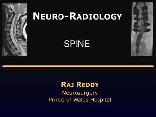
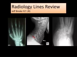
Radiology Lines Review Jeff Binder R.T. (R)
1.) yellow circle 2.)purple line 3.) red line 4. Blue lines
1.) ADI – Should be 1-5mm for children, 1-3mm for adults 2.)McGregors line – odontoid less than 8mm above this line 3.) Spinolaminar line – should be smooth curve 4.) Pre vertebral space – Top 5mm, middle 7mm, bottom 20mm
1.) Red angle 2.) yellow measurement
1.) Boehler’s angle – 28-40*, above or below this range is possible calcaneal fx 2.) Heel pad measurement – Distance between skin and calcaneus. 25<mm = acromegaly
1.) Metaphysis 2.) Physis (growth plate) 3.) Epiphysis 4.) Diaphysis (shaft) 5.) Periosteum 6.) Apophysis (attachment site for ligament or tendon)
Blue line: Iliofemoral line. Should be smooth. Fx or Slipped femoral capital epiphysis will disrupt the line Yellow line: Shenton’s line. Should be smooth curve. Fx/SCE will disrupt Purple angle: Femor Neck angle. 120*-130* is normal range. Above=Coxa Valga Below=Coxa Vara Red measurement: acetabular depth. 7-18mm average. Increased by osteomalacia, Paget’s, or RA. Decreased in congenital hip dysplasia Brown line: Skinner’s line, Fovea capitus must be below the horizontal line. Fx or SCE will elevate the line.
Blue angle: Center edge angle; 20*-40*; increased by osteomalacia, Paget’s, or RA. Decreased by join effusion Orange line: Kline’s line; altered by FX or SCE Red line: Kohler’s line; If femur head crosses the line then it is called Protrusio Acetabuli. Osteomalacia, Paget’s, and RA cause it Orange measurements: Superior (3-6mm), axial (3-7mm), and medial (4-13mm) (teardrop) distances *never average these measurements If medial joint space (teardrop) exceeds 11mm or 2mm between right and left then Waldenstrom’s sign. Common in Legg-Calve-Perthes
Blue lines: Ac joint space. Average superior and Inferior. ~3mm Red Line: Acromiohumeral joint space. 7-11mm Rotator cuff tear decreases Dislocation or joint effusion increases Yellow lines: Glenohumeral joint space. Average all 3. 4-5mm Posterior dislocation increases measurement CPPD or OA decrease measurement
RAO Position Structure, level, side
Right c4/5 IVF The first IVF is C2/3 so count down from there. This is an ROA so the right side of the neck is touching the film therefore RIGHT IVF’s will be visualized.
http://www.imaios.com/en/e-Anatomy/Spine/Spine-standard-radiographyhttp://www.imaios.com/en/e-Anatomy/Spine/Spine-standard-radiography