MS Lesion Visualization Assisted Segmentation
Explore how advanced visualization methods assist in accurately identifying MS lesions from MRI images, aiding in diagnosis and treatment planning. Overcome challenges of overlapping tissue intensities and partial volume effects through innovative segmentation techniques. Gain insights into MS progression rates and the impact on patients. Develop a more efficient and objective approach for lesion detection. Collaborate with experts to refine results and enhance accuracy. Join the project for a comprehensive understanding of multiple sclerosis visualization and segmentation.

MS Lesion Visualization Assisted Segmentation
E N D
Presentation Transcript
MS Lesion Visualization Assisted Segmentation Daniel Biediger COSC 6397 – Scientific Visualization
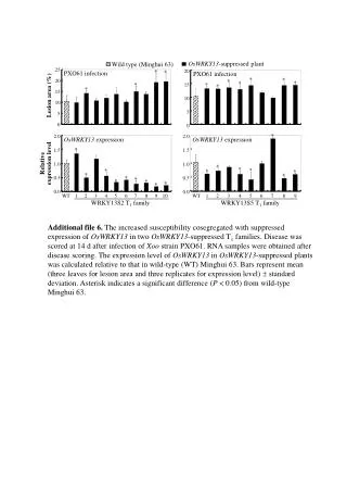
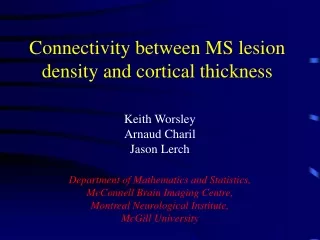
Multiple Sclerosis • Autoimmune disease of the central nervous system • Affects 2.5 million people globally, ~100 per 100,000 • Damage to the insulating myelin sheaths around brain cells • Appears as a range of cognitive and physical disability • Progresses at different rates with remission and relapse Myelin: ~40% water, ~40-50% lipids
Multiple Sclerosis • Identified in-vivo with MRI images • Normally 3 tissues: GM, WM, CSF (SG, SB, LCR) • Lesion appear hyper- or hypo-intense (bright or dark) • Reflects a difference in relaxation times for lipids/water • Detected as an outlier to the normal WM • FLAIR MRI (Fluid Attenuated Inversion Recovery) best
MS Detection Challenges • WM Lesion detection is a challenge • Overlap in intensity between tissue classes • 3D brain structure (sulcus, ventricles, multiple tissues) • Partial Volume Effects (PVE) • Artifacts from fluid/patient motion, tumors, etc • Random noise, bias field, sun spots, etc • Requires an expert to identify lesions • Tissue identification (T1-weighted MRI) • Lesion detection (FLAIR MRI) • Time consuming, difficult (multi-modality), and subjective
MS Lesion Appearance in Flair This? Is this? or This? or This?
Result Comparison FLAIR MRI Base Method Expert Me Under Segmentation Correct Segmentation Over Segmentation Results DICE = 0.65