The Nervous System rev 12-12
770 likes | 978 Vues
The Nervous System rev 12-12. Receives information and produces a meaningful, quick output. To do this, the nervous system quickly sorts through our memory bank decides the probable meaning of the input integrates the information Provides a quick response

The Nervous System rev 12-12
E N D
Presentation Transcript
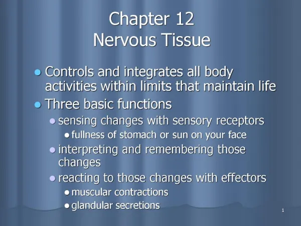
The Nervous System rev 12-12 • Receives information and produces a meaningful, quick output. • To do this, the nervous system • quickly sorts through our memory bank • decides the probable meaning of the input • integrates the information • Provides a quick response • So, the nervous system controls and integrates all other body systems and functions Nervous System HANDOUT 1
What are the characteristics of the nervous system that allow us to do this? • It must receive information from our senses. • It integrates information. -Integration is the process of taking different pieces of information from different sources, making sense of all of it at the same time, and coming up with an action plan. 3. The nervous system is fast; it can do this within tenths of a second. Nervous System HANDOUT 2
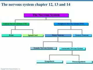
The CNS-Central Nervous System(the brain and spinal cord) • is the integrating and command center of the nervous system • It receives and interprets incoming sensory information and produces motor responses The PNS-peripheral nervous system is the part of the nervous system outside the CNS. • it contains the communication lines that link all parts of the body to the CNS Nervous System HANDOUT 3
The Periph NS consists of • 12 pairs of cranial nerves: carry impulses between brain and body • 31 pairs of spinal nerves: connect to spinal cord via dorsal and ventral roots • Dorsal root has sensory neurons and transmit information TO the cord • Ventral root has motor neurons that transmit information FROM the cord to the body Nervous System HANDOUT 4
the Peripheral NS has 2 functional subdivisions • the sensory or afferent division carries impulses TO the CNS • keeps the CNS informed of events going on inside and outside of the body • The motor or efferent division carries impulses FROM the CNS • this division enables us to respond to stimuli Nervous System HANDOUT 5
The Motor Division can be further subdivided into 2 parts: • the Somatic nervous system • Voluntary: controls voluntary and involuntary skeletal muscle movements • Motor neurons are activated either by conscious control from the brain or by an involuntary response called a reflex Nervous System HANDOUT 6
Reflex Division: • Spinal reflexes • Spinal reflexes are involuntary, automatic responses handled primarily by the spinal cord and spinal nerves And, the other part or division of the Periph NS, the Autonomic nervous system (ANS) • this division regulates involuntary activities; the activity of smooth muscles, cardiac muscles, and glands • Functions below the level of awareness • (regulates anything that occurs automatically in the body) Nervous System HANDOUT 7
Requires 2 neurons to transmit information from the CNS to a “target” cell • Preganglionic neurons: cell bodies of the first neurons lie within the CNS • Sympathetic preganglionic fibers short • Parasympathetic preganglionic fibers long • The axons of these go to postganglionic neurons which lie outside the CNS • Postganglionic axons extend to wherever in our body the target glands or organs are located Nervous System HANDOUT 8
The ANS is further sub-divided into the • SYMPATHETIC NERVOUS SYSTEM which mobilizes body systems during emergency situations. • Origin: thoracic or lumbar regions of the spinal cord • Function: releases neurotransmitter norepinephrine for fight-or-flight reaction; opposes parasympathetic division • reduces blood flow to organs that do not help with an immediate disaster Nervous System HANDOUT 9
AND the PARASYMPATHETIC NERVOUS SYSTEM(PSNS) which conserves our energy and predominates during relaxing • Origin: brain and sacral area of spinal cord (craniosacral) • Function: releases acetylcholine to relax the body; opposes sympathetic division • In most organs, the actions of the sympathetic and parasympathetic divisions have opposite effects. • The two divisions counterbalance each other’s activities to maintain homeostasis. Nervous System HANDOUT 10
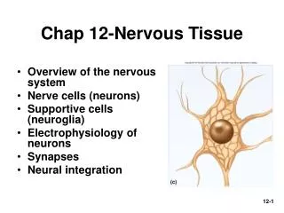
Composition of the Nervous SystemNerves, Neurons, Neuroglia The Nervous System is composed of Nerves which consists of the axons of many neurons wrapped together in a sheath of connective tissue Neurons are cells which are specialized for communication. • Classification of neurons • Sensory or Afferent neurons carry (sensory) information from receptors TO the CNS • Motor (Efferent) Neurons carry messages AWAY FROM the CNS to the muscles and the glands Nervous System HANDOUT 11
Interneurons or Association Neurons are located in the CNS and conduct impulses within the CNS. They receive information from sensory neurons, integrate the input, and then deliver the information to other neurons. • are multipolar neurons • Neuroglia (or Glia) are the supporting and protecting cells of the nervous system. We will speak more of these later. Nervous System HANDOUT 12
Neurons • All neurons have • a cell body (contains the nucleus) • an axon (long slender tube of cell membrane; specialized to carry electrical impulses) • Axons of sensory neurons originate from a dendrite • Axons of interneurons and motor neurons originate from a cone shaped area of the cell body called the axon hillock • At its other end, the axon branches into slender extensions called axon terminals and the end of this is called an axon bulb • dendrites (typically slender extensions of the cell body; receive stimuli) Nervous System HANDOUT 13
Types of Neurons Unipolar Neuronshave a single process which emerges from the cell body • this process divides into a proximal and distal branch • One branch behaves as an afferent branch and the other behaves as an efferent branch • All unipolar neurons are sensory Nervous System HANDOUT 14
Bipolar Neurons • have 2 processes emerging from a round cell body • processes extend from opposite sides of the cell body • found only in some of the special sense organs where they act as receptor cells Nervous System HANDOUT
Multipolar Neurons • have 3 or more processes • are the most common neuron type in humans and major neuron type in the CNS Nervous System HANDOUT 16
Interneurons or Association Neurons • are a multipolar neuron • located in the CNS • conduct impulses within the CNS • are the connecting link between sensory and motor neurons Nervous System HANDOUT 17
Some neurons have a myelin sheath • the myelin sheath • is a fatty wrapping around the axon which provides insulation to the axon and thus saves it energy • The process of myelination continues during childhood and is the primary reason for the increase in a child's brain size • it speeds impulse transmission by allowing a leaping pattern of transmission called saltatory conduction • The impulse jumps from one Node of Ranvier to another Nervous System HANDOUT 18
Between neighboring Schwann cells are short, uninsulated gaps called Nodes of Ranvier Nervous System HANDOUT 19 Figure 11.7a
PNS: Schwann Cell Myelin sheath CNS: Oligodendrocyte Nervous System HANDOUT
Myelin • in the peripheral nervous system is formed from Schwann Cells which wrap around the axon • It helps damaged or severed axons of peripheral nerves regenerate • in the CNS is formed by oligodendrocytes • The oligodendrocyte sheath degenerates once the axon it protects is damaged or destroyed Nervous System HANDOUT
Neuroglial cells • Provide physical support to neurons • Protection to neurons • Help maintain concentrations of chemicals in the fluid surrounding them • Neuroglial cells DO NOT generate or transmit impulses Nervous System HANDOUT 22
Examples of Neuroglial Cells: • Oligodendrocytes form the myelin sheath in the CNS • Schwann cells form the myelin sheath in the PNS • Astrocytes anchor the neurons to nearby capillaries • Ependymalcellsare ciliated cells that move cerebrospinal fluid around the brain and spinal cord • Microglia are phagocytic cells for the nervous system. They destroy microorganisms and damaged tissues near the neurons. Nervous System HANDOUT
Impulse Transmission-Summary The function of a neuron is to transmit information from one part of the body to another. • This is done in the form of electrical impulses. • An impulse arrives at the dendrite • When the impulse is strong enough, it depolarizes the membrane and the impulse is transmitted along the axon • When the impulse reaches the axon terminals, the information needs to be converted to another form of energy in order for the information to be transmitted to its target (i.e. a muscle, a gland, or another neuron) • A chemical, called a neurotransmitter, is released which allows the impulse to jump the synapse, or space, between the 2 cells Nervous System HANDOUT 24
Impulse Transmission- Definitions • Resting or Membrane Potential: a small difference in voltage across the cell membrane; the cell is normally negatively charged. • This allows the neuron to be ready to respond more quickly than it could if it were electrically neutral. • Think about a car battery. It retains a charge so that the car will start as soon as the key is turned • Unlike most body cells, neurons can alter the electrical charge across the neurolemma. The membrane potential alternates between -70 and +30 millivolts. Charge differences are controlled by the movement of sodium and potassium ions entering and leaving the neuron • Action Potential: changes in the electrical activity of the nerve of sufficient intensity to reach the threshold necessary to move an electrical impulse down the axon Nervous System HANDOUT
Threshold: the level of stimulus a neuron needs in order to fire • All or nothing phenomenon: once the threshold level is reached, the nerve transmits an impulse • Depolarization: moving the negative charge within the axon closer to zero • Na+ (sodium) moves into the cell via sodium channels which open • Na channels act as molecular amplifiers and turn small electrical signals into action potentials that can conduct for long distances Nervous System HANDOUT
Repolarization: K+ (potassium) moves out of the cell; Na+ channels close and the reversal of the membrane polarity triggers opening of the K+ channels so the K+ moves out of the cell. Loss of K+ means that the interior of the axon becomes negative again and the resting potential is restored • Refractory period: The part of the axon that has already fired is unable to fire again so the impulse must move forward (This prevents the impulse from moving backwards.) Nervous System HANDOUT
Nerve impulse transmission, explanation 2 • All cells demonstrate an unequal distribution of ions or a voltage difference across the cell membrane. This is called a MEMBRANE POTENTIAL OR A RESTING MEMBRANE POTENTIAL. It occurs when the membrane is not being stimulated. • Nerve cells and muscle cells (and some cells of glands) can produce electrochemical impulses and transmit them along their membrane when the cell is stimulated or “excited”. • When a nerve cell is stimulated sufficiently, the voltage across the membrane is changed. Nervous System HANDOUT
If the stimulus is strong enough, the impulse reaches the THRESHOLD. Once the impulse starts moving down the axon, it is called an ACTION POTENTIAL and follows an ALL OR NOTHING law. • Two ions are responsible for this: Nervous System HANDOUT
The sodium and potassium ion permeabilities of the cell membrane changes. • First, Na+ diffuses into the cell. This makes the inside of the axon more positively charged. As the cell becomes more positive, the THRESHOLD stimulus level is reached. Na+ DEPOLARIZES the membrane. • As the action potential reaches its peak, sodium channels (or gates) close and the potassium channels (or gates) open so K+ diffuses to the outside of the cell. This allowing the cell to return to its original -70 mV charge or its resting potential. The Na+--K+ pump helps to maintain this ‘normal’ charge across the cell membrane. Nervous System HANDOUT
The change of electrical current occurs in a small portion of the cell membrane at any one time. This allows the charge to move forward in a step-like progression. • Once activation of a cell has begun, the membrane is generally insensitive to any further stimulation. This is called the REFRACTORY PERIOD. Near the end of the activation impulse, the cell can be activated but only if a stimulus is much stronger than normal. Nervous System HANDOUT
PART IIThe Brain and the Spinal Cord rev 12-12 The CNS controls and processes all information received by the body. • Protection of the CNS • Bone protects it from physical injury • the brain is covered by the skull • the spinal cord is surrounded by the vertebrae Nervous System HANDOUT 32
The Brain • The brain is the central command center of the body • Receives information in the form of action potentials from various nerves and the spinal cord, integrates it and generates the appropriate response. • 3 major anatomical and functional divisions of the brain have been identified: Nervous System HANDOUT
Brain: Major Divisions • Hindbrain: coordinates basic, automatic, and vital functions • Midbrain: helps coordinate muscle groups and responses to sight and sound • Forebrain: receives and integrates sensory input from the external and internal environment and determines most of our complex behavior Nervous System HANDOUT
Hindbrain: movement and automatic functionsMedulla, Pons, Cerebellum • Connected to the spinal cord • Is the oldest, most primitive brain division Medulla Oblongata: controls automatic functions of internal organs • cardiovascular center- regulates heart rate and blood pressure • respiratory center-controls rate and depth of breathing • other centers which coordinates reflexes such as coughing, vomiting, swallowing, and sneezing Nervous System HANDOUT
All information passing between the spinal cord and the higher areas of the brain must pass through the medulla • Motor nerves from one side of the forebrain cross over to the other side of the body in the medulla • Left side of brain controls right side of body • Right side of brain controls left side of body Nervous System HANDOUT
Pons • Aids information flow • connects the higher brain centers and the spinal cord • its respiratory nuclei work with the respiratory centers of the medulla in regulating respiration • coordinates the information flow between the cerebellum and higher brain centers Nervous System HANDOUT
Cerebellum • Coordinates basic movements below the level of conscious control • Stores and produces whole sequences of skilled movements i.e. driving • Receives sensory input from many sources—joints, muscles, balance and position receptors in the ear, and visual receptors • excessive alcohol disrupts normal functioning of the cerebellum; this is why drunk people lose the ability to coordinate their movements including walking a straight line Nervous System HANDOUT
The Midbrain—functions relate to vision and hearing • Visual and auditory sensory inputs pass through the midbrain before being relayed to higher brain centers • Coordinates movements of the head in response to vision and hearing • controls movement of the eyes and pupil size • monitors the unconscious movement of skeletal muscles so their actions are smooth and coordinated • The reticular formation extends through the medulla, the pons, and the midbrain. • works with the cerebellum to coordinate muscle activity to maintain posture, balance and muscle tone • The Reticular Activating System, within the reticular formation, is responsible for maintaining our level of wakefulness Nervous System HANDOUT
The forebrain and diencephalon: emotions and conscious thought Important areas are the cerebrum, thalamus, hypothalamus, and limbic system • Also includes 2 glands: the pineal gland and the pituitary gland • Determines our most complex behavior including emotions and conscious thought. Hypothalamus and Thalamus maintain homeostasis and process information • Hypothalamus is a small region at the base of the forebrain that coordinates some automatic functions including regulating homeostasis due to monitoring of sensory signals • Has a hunger center and a thirst center • Also controls the pituitary gland Nervous System HANDOUT
Thalamus: located just above the hypothalamus • Is primarily a receiving, processing and transfer center; it sends signals to the cerebrum to be interpreted. (We become conscious of information when it arrives at the thalamus, but only after information arrives at the cerebrum are we aware of which part of the body is experiencing the information. • Limbic Systemis a group of neuronal pathways which connect parts of the thalamus, hypothalamus and cerebrum. • involved in emotions and memory. Nervous System HANDOUT
Cerebrumdeals with higher functions and is most highly developed • is divided into left and right “cerebral hemispheres” by the longitudinal fissure • each hemisphere controls the opposite side of the body • the left hemisphere controls the right side of the body • the right hemisphere controls the left side of the body Nervous System HANDOUT
In the middle of the hemispheres is the corpus callosum which joins the 2 hemispheres and enables them to communicate and share information • Below the corpus callosum in each hemisphere are the lateral ventricles which secrete CSF • The outer layer of the cerebrum is called the cerebral cortex and is primarily gray matter • The inner portion of the cerebrum is primarily white matter Nervous System HANDOUT
surface tissue of the cerebrum is covered with sulci (grooves) and gyri (ridges) which increase the surface area for information exchange Nervous System HANDOUT
Each hemisphere is further divided into 4 lobes: the frontal, parietal, temporal, and occipital lobes • all 4 lobes are involved in memory storage Nervous System HANDOUT
The frontal lobes initiate motor activity and are responsible for speech, conscious thought, and personality. • these lobes may be further divided into the prefontal lobes or cortex (the most forward area of these lobes) which are the intellectual center • intelligence, motivation, personality, abstract reasoning, judgment, planning, love, concern for others • the premotor cortex • skilled repetitive activities (keyboarding/typing) and conditioned reflexes (Pavlov’s dog) Nervous System HANDOUT
the primary motor cortex which initiates voluntary motor activity of the arms, legs, trunk and face • In the dominant hemisphere only is our primary speech center Nervous System HANDOUT
The parietal lobes house the somatosensory cortex • interpret sensory information from the skin and from proprioceptors in the muscles and joints. • This allows us to identify the region of our body being stimulated without looking • Stereognosis: integrate different sensory inputs to allow us to interpret sensory information i.e. reaching into your pocket and being able to interpret the coins in it without using visual cues. Nervous System HANDOUT
The occipital lobes house the primary visual cortex and the visual association area • association areas allow us to interpret/understand information • The temporal lobes interprets auditory information and is responsible for perceptual judgment Nervous System HANDOUT
Cranial Nerves • Twelve pairs of cranial nerves arise from the brain • They have sensory, motor, or both sensory and motor functions • Each nerve is identified by a number (I through XII) and a name Nervous System HANDOUT