Approach to the patient with Adrenal Incidentalomas
1. Approach to the patient with Adrenal Incidentalomas. SH. Alamdari , MD Associate professor of Internal Medicine, Endocrinology & Metabolism Chief, Department of Internal Medicine Shahid Beheshti University of Medical Sciences. Increasing prevalence.


Approach to the patient with Adrenal Incidentalomas
E N D
Presentation Transcript
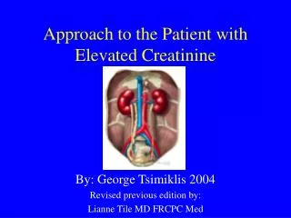
Approach to the patient with Adrenal Incidentalomas SH. Alamdari, MD Associate professor of Internal Medicine, Endocrinology & Metabolism Chief, Department of Internal Medicine ShahidBeheshti University of Medical Sciences
Increasing prevalence • The prevalence of incidentally discovered adrenal masses (also referred to as adrenal incidentalomas or adrenal nodules) has grown substantially with the increased use of cross-sectional imaging such as computed tomography (CT) and magnetic resonance imaging (MRI).
Clinical Scenarios • Adrenal masses may be discovered incidentally during emergency room visits or during the course of ambulatory or hospitalization-related imaging of the abdomen or chest. • Increasingly, adrenal masses may also be discovered during the course of surveillance imaging associated with cancer care, though these findings are not truly incidental.
Aim of this symposium • Though adrenal masses, or adrenal hyperplasia or thickening, of any size may have clinical relevance, this symposium will focus on the evaluation of incidentally discovered adrenal masses greater than or equal to 1 cm in size.
True prevalence • The true prevalence of adrenal masses is not known; rather, estimates are extrapolated from limited observations. • The prevalence of incidental adrenal masses based on 25 postmortem series is about 6% (range, 1.1 to 32%). • The prevalence of incidentally discovered adrenal masses using high-resolution CT scans is slightly lower, at approximately 4%and generally observed to be higher with older age
Why increase in the incidental discovery? • Given that the annual frequency of CT and MRI imaging studies exceeds 100 million per year in the United States, a statistic that has been steadily increasing in the last decade, the absolute number of annually discovered adrenal masses is expected to be large (10). • As modern medicine continues to increase its reliance on abdominal imaging techniques, there will likely be a parallel increase in the incidental discovery of adrenal masses.
Risk factors for developing adrenal masses • Unfortunately, our understanding of risk factors for developing adrenal masses, and our ability to predict either their presence and/or outcomes after diagnosis, is limited. • Clinicians will be faced with the challenge of evaluating these adrenal masses to ensure that they do not represent health hazards or harbingers of future risk.
The two fundamental questions • The two fundamental questions each clinician is faced with when an adrenal mass is identified are: • 1. Does this adrenal mass represent a malignancy? • 2. Is there evidence for clinically relevant and autonomous adrenal hormone excess?
Other specific questions • Other specific questions may also arise in the context of each patient’s specific situation, for example: • 3. Would a biopsy of the adrenal mass aid with the diagnosis, management, or prognosis? • 4. Is there an indication for surgical or medical treatment? • 5. Is there any indication for longitudinal surveillance with imaging and/or biochemical testing? If so, how frequently and for what duration?
Five patient scenarios • Herein, we will review the approach to answering these key questions by providing five patient scenarios, each selected to highlight a unique and relatively characteristic presentation that clinicians may encounter.
Clinical practice recommendations • Further, clinical practice recommendations should rely on the highest grades of evidence; however, since there are few randomized controlled trials and longitudinal cohort studies to dictate the most efficacious and cost-effective evidence-based approach to managing incidentally discovered adrenal masses, this review relies on lower grades of evidence (some cohort studies, but mostly retrospective and cross-sectional studies, anecdotal observations, and expert opinion).
Guiding framework for clinical care • It is with these limitations that these recommendations are presented as a guiding framework for clinical care that will undoubtedly evolve in the future, and with the knowledge that each clinician must ultimately rely on their personal judgment, and the informed opinion of their individual patients, when pursuing collective decision-making.
SCENARIO 1 • A 50-year-old woman presented to the emergency department with worsening left lower-quadrant pain. • An abdominal CT scan with intravenous contrast was performed to evaluate for diverticulitis. • No gastrointestinal abnormalities were seen; however, a right adrenal mass was noted. • She was seen by her primary care doctor 1 month later.
SCENARIO 1 • She was normotensive, normokalemic, and had no signs or symptoms of Cushing syndrome (CS). • A repeat CT scan without intravenous contrast was ordered to specifically evaluate the right adrenal mass, which was noted to be 1.0 cm in size and described as being round and homogenous with an unenhanced attenuation of 8 Hounsfield units (HU) • The left adrenal gland was normal. • A 1-mg dexamethasone suppression test was performed, yielding a morning cortisol of 1.3 μg/dL.
SCENARIO 1 Questions • Q 1: Does this adrenal mass represent a malignancy? • Q 2: Is there evidence for clinically relevant and autonomous adrenal hormone excess? • Q 3: Would a biopsy of the adrenal mass help with diagnosis or management? • Q 4: Is there an indication for surgical or medical treatment? • Q 5: Is there any indication for longitudinal surveillance with imaging and/or biochemical testing? If so, how frequently and for what duration?
SCENARIO 1 Q 1: Does this adrenal mass represent a malignancy? • No, the radiographic characteristics of this patient’s incidentally discovered right adrenal mass are suggestive of a benign adrenocortical adenoma.
SCENARIO 1Q 1: Does this adrenal mass represent a malignancy? • Adrenal masses may be benign or malignant and hormonally functional or nonfunctional. • Most incidentally discovered adrenal masses will represent a benign entity, of which the most common etiology is by far an adrenocortical adenoma
SCENARIO 1Q 1: Does this adrenal mass represent a malignancy? • Although primary adrenal malignancy (adrenocortical carcinoma) is very rare, • the prevalence of metastases to the adrenal from extra-adrenal malignancies is not uncommon and • may be increasing as cancer survival improves.
SCENARIO 1Q 1: Does this adrenal mass represent a malignancy? • The determination of malignant potential is usually a function of the radiographic appearance and size of the adrenal mass. • Adrenocortical adenomas are usually lipid-rich, a feature that classically manifests as a low attenuation on unenhanced CT (<10 HU) and/or the presence of chemical shift on MRI
SCENARIO 1 Q 1: Does this adrenal mass represent a malignancy? • In cases when the unenhanced CT attenuation is not characteristically lipid rich (>10 HU), a benign lipid-poor adrenocortical adenoma is statistically still the most likely diagnosis. • In these instances, in- and out-of-phase imaging on MRI can be performed to obtain additional information, but may provide similar information, since chemical shift also assesses lipid content.
SCENARIO 1Q 1: Does this adrenal mass represent a malignancy? • Alternatively, CT imaging with a contrast washout protocol may provide additional diagnostic value. • The most widely used washout protocol entails obtaining an unenhanced CT image at base line, followed by administration of intravenous contrast and repeat imaging at 1 minute and again at 15 minutes. • Benign and lipid-rich adenomas are not contrast avid and will usually exhibit a >60% absolute washout of contrast after 15 minutes, with a >40% relative washout
SCENARIO 1Q 1: Does this adrenal mass represent a malignancy? • Most data and experience with characterizing adrenal masses has been with CT; however, CT imaging exposes patients to radiation. • MRI is an alternative with no radiation exposure; however, the procedure takes longer than CT and is more costly. • It is important to note that high-quality evidence to support the most effective imaging approach and modality is lacking, and therefore, these recommendations are based on expert opinion and reasonable clinical judgement
SCENARIO 1Q 1: Does this adrenal mass represent a malignancy? • Other imaging characteristics that should raise concern for a potential malignancy include: • fluorodeoxyglucose(FDG) avidity on positron emission tomography, • irregular (not round) shape, • calcifications, and • a rapid rate of growth if serial imaging is available
SCENARIO 1Q 1: Does this adrenal mass represent a malignancy? • Finally, large size (>4 cm) is sometimes used as a crude indicator of a potentially malignant or concerning lesion; however, it should be noted that malignant lesions may present as small in size, and benign lesions may present as large masses • Therefore, other radiographic characteristics are usually more specific indicators than size alone.
Q 2: Is there evidence for clinically relevant and autonomous adrenal hormone excess?
SCENARIO 1Q 2: Is there evidence for clinically relevant and autonomous adrenal hormone excess? • This patient exhibits no evidence of clinically relevant hypercortisolism. • There is no strong indication to screen for primary aldosteronism in the absence of hypertension or hypokalemia, and the imaging characteristics are not suggestive of pheochromocytoma.
SCENARIO 1Q 2: Is there evidence for clinically relevant and autonomous adrenal hormone excess? • Even when radiographic characteristics support an adrenal mass as being benign, a biochemical evaluation for adrenal hormone excess should be considered. • Since most adrenal masses are detected incidentally, these patients usually do not exhibit symptoms or signs of an overt syndrome of adrenal hormone excess such as CS, primary aldosteronism, or pheochromocytoma.
SCENARIO 1Q 2: Is there evidence for clinically relevant and autonomous adrenal hormone excess? • However, even mild or autonomous hypercortisolismcan increase the risk for future cardiometabolic and skeletal risk • Accordingly, it is generally recommended that all patients with an adrenal mass be evaluated with an overnight 1-mg dexamethasone suppression test, where postdexamethasone morning cortisol values ≤1.8 μg/dL are considered to be “normal” or indicative of “nonfunctional” status
SCENARIO 1Q 2: Is there evidence for clinically relevant and autonomous adrenal hormone excess? • Patients with hypertension and/or hypokalemia should be screened for primary aldosteronism, and screening for pheochromocytoma should be considered in all patients with a lipidpoor adrenal mass, since it is important to detect pheochromocytoma prior to the development of overt adrenergic symptoms
SCENARIO 1Q 2: Is there evidence for clinically relevant and autonomous adrenal hormone excess? • Patients with a homogenous, non– contrast avid, and lipid-rich adrenal mass (<10 HU) do not require biochemical evaluation for pheochromocytomabecause the case detection in such scenarios is close to zero and complicated by a relatively high rate of false-positive biochemical testing
Q 3: Would a biopsy of the adrenal mass help with diagnosis or management?
SCENARIO 1 Q 3: Would a biopsy of the adrenal mass help with diagnosis or management? • There is no role for an adrenal biopsy in this case. The role of adrenal mass biopsy is limited. • A biopsy should be considered when: • an extra-adrenal metastasis or an infection are the suspected etiology of the adrenal mass(es) and • when the information from cytology is likely to influence management.
SCENARIO 1Q 3: Would a biopsy of the adrenal mass help with diagnosis or management? • Extra-adrenal metastases should be suspected when: • a patient has a history of nonadrenal cancer, • when there are bilateral adrenal masses, and • the radiographic appearance of the masses is dense and vascular and/or with an irregular shape (classically, neither round nor with smooth contour). • In these situations, a biopsy can determine the type and stage of the primary cancer, which may influence subsequent management.
SCENARIO 1Q 3: Would a biopsy of the adrenal mass help with diagnosis or management? • A biopsy may also be considered when an infiltrative infection to the adrenal glands is suspected, such as by Mycobacterium or fungal organisms, and there are no other means to confirm the diagnosis. • Patients with infectious adrenal infiltrations are often partially immunocompromised or had lived or traveled in potentially endemic regions. • A biopsy in this situation may inform therapy towards specific antimicrobials rather than surgical procedures.
SCENARIO 1Q 3: Would a biopsy of the adrenal mass help with diagnosis or management? • A biopsy is not recommended to distinguish a primary adrenocortical adenoma from a carcinoma; biopsy in these situations may be inaccurate and/or falsely reassuring due to sampling bias, and there is a theoretical risk of tumor seeding the needle track • When concern for a primary adrenal malignancy is raised based on radiographic imaging characteristics, a cancer-focused surgery should be strongly considered for diagnosis, prognosis/ staging, and treatment.
SCENARIO 1Q 3: Would a biopsy of the adrenal mass help with diagnosis or management? • Infrequently, a suspected adrenal malignancy is deemed to be surgically unresectable, yet confirmation of the malignancy via histopathology is necessary to determine appropriate medical therapy (i.e., chemotherapy). • In this circumstance, a biopsy of a likely malignant mass may be considered.
SCENARIO 1 Q 3: Would a biopsy of the adrenal mass help with diagnosis or management? • Biopsy is contra-indicated in any mass which could represent a pheochromocytoma due to concern of precipitating a catecholamine crisis; rather, biochemical diagnosis, pre-operative adrenergic receptor blockade, and surgery should be pursued.
Q 4: Is there an indication for surgical or medical treatment?
SCENARIO 1 Q 4: Is there an indication for surgical or medical treatment? • There is no indication for surgical or medical treatment in this case, since the adrenal mass is neither suspicious for malignancy nor hormonally active.
SCENARIO 1Q 4: Is there an indication for surgical or medical treatment? • History and physical examination should first be conducted to assess suspicion for an obvious syndrome of adrenal hormone excess, followed by biochemical testing for evidence of adrenal hormone excess. • If hormone excess is confirmed, localization of the source and surgical resection should be strongly considered
SCENARIO 1Q 4: Is there an indication for surgical or medical treatment? • Most patients with an incidentally discovered adrenal mass are asymptomatic and do not exhibit an obvious syndrome of adrenal hormone excess and have normal values on biochemical screening. • In these instances, the radiographic characteristics of the adrenal mass dictate subsequent management.
SCENARIO 1Q 4: Is there an indication for surgical or medical treatment? • Adrenal masses that display suspicious or concerning features for a potential malignancy can be further characterized using alternative imaging modalities if needed, and if unilateral, surgically removed. • When there is uncertainty regarding malignant potential or ambiguous characterization that prevent firm decision making, repeated imaging can be performed in 3 to 6 months to reassess for progressive radiographic or biochemical features that may confirm suspicions of a potential malignancy