Download
1 / 52
520 likes | 546 Vues
Understand the intricate world of iron metabolism from a renowned expert in the field, exploring the journey of iron from essentiality to toxicity. Learn about iron's roles, absorption mechanisms, storage, and regulatory hormones for optimal health.

E N D
IRON: FROM DEFICIENCY TO OVERLOAD DR. MOHAMED EL FAKI OSMAN, MD Assistant Professor & Consultant Paediatric Haematologist/Oncologist Department of Paediatrics & King Khalid University Hospital College of Medicine King Saud University Riyadh, KSA
بسم الله الرحمن الرحيـــم • " لَقَدْ أَرْسَلْنَا رُسُلَنَا بِالْبَيِّنَاتِ وَأَنْزَلْنَا مَعَهُمُ الْكِتَابَ وَالْمِيزَانَ لِيَقُومَ النَّاسُ بِالْقِسْطِ وَأَنْزَلْنَا الْحَدِيدَ فِيهِ بَأْسٌ شَدِيدٌ وَمَنَافِعُ لِلنَّاسِ وَلِيَعْلَمَ اللَّهُ مَنْ يَنْصُرُهُ وَرُسُلَهُ بِالْغَيْبِ إِنَّ اللَّهَ قَوِيٌّ عَزِيزٌ " الحديد (25)
Iron lacks the glitter of gold and the sparkle of silver but outshines both in biologic importance* *Nancy C Andrews, in Hematology of Infancy and Childhood
Iron Metabolism/Facts • Iron exists in two stable oxidation states: • Ferrous (Fe+2) • Ferric (Fe+3) • Can act as a redox catalyst i.e. reversibly donating or accepting electrons
Iron Metabolism/Facts • Adult body iron contents is 3-5 gm (35-45mg/kg) • 75% (60% - 80%) in Hb • 25% stored in liver, RES macrophages, in haem containing proteins e.g myoglobin, cytochrome P-450, Myeloperoxidase…etc.
Iron Metabolism/Facts • Haemoglobin structure • Hb molecule is a tetramer (4 polypeptide chains) • 2 α chains (141 άά) • 2 β chains (146 ά ά) • Each chain attached to a prosthetic group (haem) • Haem→ protoporphyrin IX + iron molecule (Fe+2)
Iron Metabolism/Homeostasis • The daily requirement 10-20mg • The daily absorption 1-2mg(10%) • The daily excretion 1-2mg • The daily utilization in bone marrow for Erythropoiesis 20mg • Excess iron stored in liver and RES
Iron Metabolism/Homeostasis • Iron Absorption • Mostly in the upper GIT • Haem iron is readily absorbed • Non-haem iron (inorganic) → duodenum entrocyte as Ferric (Fe+3) brush border Ferrous (Fe+2) Ferric reductase DMT1* entrocyte *DMT-1 = Divalent Metal Transporter 1
Iron Metabolism/Homeostasis • Duodenal entrocyte iron • Retained by the cell and lost when the cell dies and sloughed (excretion) • Transported through cell to enter the body by → Ferroportin • Binds to Transferrin in plasma
Iron Metabolism/Homeostasis • Role of Transferrin • The main iron carrying glycoprotein in plasma • Synthesized in the liver (sertoli cells, oligodendrocytes) • Renders iron more soluble • Prevents iron-mediated free radical toxicity • Facilitates transport into the cells
Iron Metabolism/Homeostasis • Role of transferrin • 1 apotransferrin molecule + 2 iron atoms → differic transferrin → transferrin receptor on cell surface → iron release to the cell. • Non transferrin bound iron (NTBI) is toxic to tissues • Mitochondria for Hb synthesis • Ferritin for storage
Iron Metabolism/Homeostasis • Ferritin • Complex protein sub units • Sphere with a central cavity • Several thousands atoms of crystalline iron • Metabolically inactive and non-toxic • Mostly intracellular • Measurable amount in serum • Circulating Ferritin is iron-poor
Iron Metabolism/Homeostasis • Role of Hepcidin: • Hepcidin is the main hormone regulating iron homeostasis(2000) • 25- α a glycoprotein synthesized in the liver • Binds to ferriportin leading to its degradation → suppression of iron release from intestine and from macrophages, stable iron levels
Iron Metabolism/Homeostasis • Role of Hepcidin • Levels increase in : • Inflammation/infection • Iron overload • Reduced in: • Hypoxia • Anaemia
Iron Metabolism/Homeostasis • Hemosiderin • Ferritin molecules aggregate → clusters • Engulfed and degraded by lysosomes • Denaturated protein and lipid interspered with iron oxide molecules
Iron Metabolism/Homeostasis • Non-Transferrin-Bound Iron • Level increases in complete transferrin saturation states eg hypotransferrinaemia and in iron overload • Weakly complexes with albumin, citrate, amino acids and sugars • Preferentially taken by non-hematopoetic cells e.g. liver, endocrine organs, kidneys and heart • Highly toxic to cells • Potentiates formations of free radicals → cell membrane damage and death
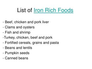
Extremes of Iron Balance • 1. Iron deficiency • Causes • Inadequate absorption • Poor bio availability (absorption of haem fe >Fe2+ > Fe3+) • Antacid therapy/high gastric ph • Bran, tannins, phytates, starch • Other metals e.g. cobalt, lead • Loss /dysfunction of absorptive enterocytes • Insufficient iron stores • Bleeding; GIT, urinary, pulmonary • Inadequate presentation to erythroid precursors • Atransferrinemia • Anti-transferrin receptors antibodies
Consequences of Iron Deficiency • Three stages of iron deficiency • Prelatent iron deficiency (Iron Depletion) • Reduction of iron stores • No change in haematocrit • No change in serum iron level • Detected by low serum ferritin level
Consequences of Iron Deficiency • Three stages of iron deficiency (Continue) • Latent Iron Deficiency (Iron Deficient Erythropoiesis) • Shortage of iron available for Hb synthesis • No change in haematocrit • Low serum iron and ferritin levels • Increase total iron binding capacity (TIBC) • Detected by low early morning transferrin saturation • Increase in free erythrocyte zinc protoporphyrin
Consequences of Iron Deficiency • Three stages of iron deficiency (Continue) • Iron deficiency anaemia • Serve hypo chromic microcytic anaemia • Low serum iron levels • Low serum ferritin levels • Low transferrin saturation • High TIBC
Urinary hepcidin level as an early predictor of iron deficiency in children: A case control studyMohammed Sanad1* and Amal F Gharib2 • 25 normal children • 25 children with stage I ID • 25 children with stage 2 ID • 25 children with stage 3 ID
Urinary hepcidin level as an early predictor of iron deficiency in children: A case control studyMohammed Sanad1* and Amal F Gharib2 • Urinary hepcidin level is normal in all 25 normal children (control group) • Urinary hepcidin levels were low in all three stages of iron deficiency • More significantly low with the progression of iron deficiency
Signs and symptoms of ID • No symptoms • Findings of anaemia; weakness, fatigue, palpitations, lightheadedness • Epithelial changes; angular stomatitis, glossitis, smooth tongue,gastric atrophy, koilonychia (spoon nails) • Plummer-Vinson syndrome (oesophageal web syndrome) • Pica (compulsive consumption of non-nutritive materials)
Diagnosis; • Bone marrow smear shows no stainable iron is the only definitive diagnosis. • Low serum iron level • Low ferritin level • High TIBC. are considered diagnostic • Transferrin saturation should be less than 10%
Treatment of ID • Treat the cause? • Oral iron supplementation: - Ferrous sulfate is recommended 6mg/kg/day - Ascorbic acid may enhance intestinal absorption
Response to iron therapy • Reticulocytosis by day 4 • Maximum by 7-10 days • Hb may rise significantly by 2-4 weeks • Complete Hb correction by 6-8 weeks • Continue 2-3 months after correction of the anaemia to replenish the body stores
Causes of poor response to iron therapy • Noncompliance • Ongoing blood loss • Insufficient duration of therapy • High gastric pH; antacids • Inhibitors of iron absorption/utilization • Lead • Chronic inflammation • Incorrect diagnosis • Thalassemia • Sideoblastic anaemia
Parenteral iron replacement • Three forms: • Iron dextran • Iron gluconate • Iron sucrose • Indications: • Oral iron is poorly tolerated • Rapid replacement of iron stores • GIT absorption is compromised • Erythropoietin therapy is needed e.g. patients on dialysis
Iron replacement in infants • Term babies has iron stores adequate for about 6 months • Preterm babies deplete iron stores by third-fourth month • Human milk contains high amount of iron which decreases by the 5th month of lactation • Iron from human milk is 20-50% absorbed
Recommendations • Infants who are exclusively breast fed; give iron 1mg/kg/day after 6 months • Non-breast milk fed infants should be on iron-supplemental formula (12 mg/L) till the end of the first year • Start iron-enriched cereals with solid food • Avoid cow’s milk during the first year. May chelate iron and may cause GIT hemorrhage
Extremes of Iron Balance • 2. Iron overload • Primary (hereditary) • Secondary (acquired)
Secondary iron overload • Blood/PRBC transfusion • Erythroid hyperplasia • B+-thalassemia (Thalassemia intermedia) • X-linked sideroblastic anaemia • both condition lead to increase iron absorption • MDS
Primary Iron Overload • Hepcidin feedback mechanism
Primary Iron Overload (continue) • Levels of circulating iron control hepatic hepcidin production • Hepcidin production increases at times of high iron levels → degradation of ferriportin → less iron absorption • Hepcidin deficiency →increase iron level leading to haemochromatosis
Consequences of Iron Overload • Iron reacts with reactive oxygen intermediates; oxygen superoxide (O2) and hydrogen peroxide (H2O2) → free radicals, hydroxyl radicals (HO) → severe tissue damage
Consequences of Iron Overload (continue) • Liver → hepatomegaly (early) → fibrosis and micronodular cirrhosis. Diagnosed by MRI, liver biopsy • Heart → congestive cardiomyopathy, rarely pericarditis. Conduction defects → sudden death.
Consequences of Iron Overload (continue) • Endocrine organs • Endocrine pancreatic dysfunction → diabetes mellitus • Pituitary dysfunction → several endocrine problems e.g. delayed sexual maturation - Hypoparathyroidism • Thyroid is usually preserved • Skin and joints
Laboratory findings • High iron • High ferritin • Low TIBC • Transferrin saturation is increased • MRI/liver biopsy are diagnostic
Management of primary haemochromatosis • Venesection • Induction phase; 7ml/kg weekly • Follow up by ferritin level aiming to < 50 µg/d and Hb = 11gm/dl • Avoid vitamin C • ? Oral chelation • Hepcidin correction may be a future therapy
Iron Chelation Bind non-transferrin-bound iron (NTBI) → soluble → urinary excretion/intestinal