Download
1 / 42
450 likes | 763 Vues
Dr. Hassan Shaibah. The Human Body: An Orientation. Midbrain@gmail.com Ext.4011. An Overview of Anatomy. Anatomy – the study of the structure of the human body Physiology – the study of body function. An Overview of Anatomy. Anatomical terminology – based on ancient Greek or Latin

E N D
Dr. Hassan Shaibah The Human Body:An Orientation Midbrain@gmail.comExt.4011
An Overview of Anatomy • Anatomy – the study of the structure of the human body • Physiology – the study of body function
An Overview of Anatomy • Anatomical terminology – based on ancient Greek or Latin • Provides standard nomenclature worldwide • Branches of anatomy • Gross anatomy • Microscopic anatomy (histology)
An Overview of Anatomy • Other branches of anatomy • Developmental anatomy • Embryology • Pathological anatomy (pathology) • Radiographic anatomy • Functional morphology
The Hierarchy of Structural Organization • Chemical Level – atoms form molecules • Cellular level – cells and their functional subunits • Tissue level – a group of cells performing a common function
The Hierarchy of Structural Organization • Organ level – a discrete structure made up of more than one tissue • Organ system – organs working together for a common purpose • Organismal level– the result of all simpler levels working in unison
The Hierarchy of Structural Organization Figure 1.1
The Body Organ Systems • Integumentary • Skeletal • Muscular • Nervous • Endocrine • Cardiovascular • Lymphatic • Respiratory • Digestive • Urinary • Reproductive
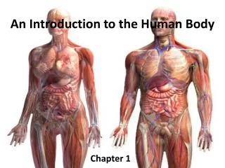
Gross Anatomy – An Introduction • Anatomical position – a common visual reference point • Person stands erect with feet together and eyes forward • Palms face anteriorly with the thumbs pointed away from the body • Directional terminology – refers to the body in anatomical position
Gross Anatomy – An Introduction Figure 1.3
Gross Anatomy – An Introduction • Directional terms • Regional terms – names of specific body areas • Axial region – the main axis of the body • Appendicular region – the limbs
Orientation and Directional Terms Table 1.1; Superior - Anterior
Orientation and Directional Terms Table 1.1; Posterior – Intermediate
Orientation and Directional Terms Table 1.1; Proximal – Deep
Regional Terms Figure 1.4a
Regional Terms Figure 1.4b
Body Planes and Sections • Coronal (frontal) plane • Lies vertically and divides body into anterior and posterior parts • Median (midsagittal) plane • Specific sagittal plane that lies vertically in the midline
Body Planes and Sections • Transverse plane • Runs horizontally – divides body into superior and inferior parts Figure 1.5
Basic Human Body Plan and Structures Shared with all Vertebrates Figure 1.7c
Organization of the Human Body • Axial, appendicular portions of body • Body cavities • Dorsal cavity • Cranial, vertebral canal • Ventral cavity • Thoracic • Abdomenopelvic • Separated by diaphragm
Body Cavities and Membranes Figure 1.8a
Body Cavities and Membranes Figure 1.8b
Body Cavities and Membranes • Serous cavities – a slit-like space lined by a serous membrane • Pleura, pericardium, and peritoneum • Parietal serosa – outer wall of the cavity • Visceral serosa – covers visceral organs
Serous Membranes • Line thoracic and abdomenopelvic cavities • Line body wall and fold back over organs • Secrete water, salts; slippery • Parietal, visceral layers • Thoracic membranes • Pleural membranes with space, fluid between layers • Pericardial membranes with space, fluid between • Abdominal membranes • Peritoneal membranes with space (peritoneal cavity between layers)
Body Cavities and Membranes Figure 1.9a, b
Body Cavities and Membranes Figure 1.9c
Body Cavities and Membranes Figure 1.9d
Body Cavities and Membranes • Other cavities • Oral cavity • Nasal cavity • Orbital cavities • Middle ear cavities • Synovial cavities
Other Body Cavities Figure 1.10
Abdominal Regions and Quadrants • Abdominal regions – divides abdomen into nine regions • Abdominal quadrants – divides abdomen into four quadrants
Abdominal Regions Figure 1.11a, b
Abdominal Quadrants Figure 1.12
Microscopic Anatomy • Microscopy – examining small structures through a microscope • Light microscopy – illuminates tissue with a beam of light (lower magnification) • Electron microscopy – uses beams of electrons (higher magnification)
Microscopic Anatomy • Preparing human tissue for microscopy • Specimen is fixed (preserved) and sectioned • Specimen is stained to distinguish anatomical structures • Acidic stain – negatively charged dye molecules • Basic stain – positively charged dye molecules
Microscopic Anatomy • Scanning electron microscopy • Heavy metal salt stain – deflects electrons in the beam to different extents • Artifacts • Minor distortions of preserved tissues • Not exactly like living tissues and organs
Clinical Anatomy – An Introduction to Medical Imaging Techniques • X ray – electromagnetic waves of very short length • Best for visualizing bones and abnormal dense structures
Clinical Anatomy – An Introduction to Medical Imaging Techniques • Variations of X ray • Fluoroscope – images are viewed on a fluorescent screen • Allows viewing of internal organs as they move • Cineradiography – uses X-ray cinema film to record organ movements
Advanced X-Ray Techniques • Computed (axial) tomography (CT or CAT) – takes successive X rays around a person's full circumference • Translates recorded information into a detailed picture of the body section
Advanced X-Ray Techniques • Digital subtraction angiography imaging (DSA) – provides an unobstructed view of small arteries • DSA is often used to identify blockages of arteries that supply the heart or brain
Advanced X-Ray Techniques • Positron emission tomography (PET) – forms images by detecting radioactive isotopes injected into the body • Sonography (ultrasound imaging) – body is probed with pulses of high-frequency sound waves that echo off the body's tissues • Imaging technique used to determine the age of a developing fetus
Advanced X-Ray Techniques • Magnetic resonance imaging (MRI) – produces high-quality images of soft tissues • Distinguishes body tissues based on relative water content