Hemoglobin A 2
Practical Hematology Lab. - LAB 6 -. Hemoglobin A 2. Introduction. HBA 2 is a protein which in humans is encoded by the HBA 2 gene. Hemoglobin A 2 is a normal variant of hemoglobin A that consists of two alpha and two delta chains and is found in small quantity in normal human blood.


Hemoglobin A 2
E N D
Presentation Transcript
Practical Hematology Lab - LAB 6 - Hemoglobin A2
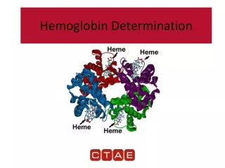
Introduction • HBA2 is a protein which in humans is encoded by the HBA2 gene. • Hemoglobin A2 is a normal variant of hemoglobin A that consists of two alpha and two delta chains and is found in small quantity in normal human blood. • Normal value of HBA2 in the adult is ( 1.8 – 3.5%).
HbA2 elevated up to 8% indicates: • β. Thalassemia trait • Homozygous Thalassemia • Decreased level may be found in: • iron deficiency anemia. • Hb H disease. • hereditary persistence of Hb F. • fibroblastic anemia. • carriers of α Thalassemia.
HbA2 test – Anion Exchange Chromatography • The anion exchange micro chromatography procedure is an accurate and easily performed method for HbA2 Quantization. • Principle: • A hemolysate is prepared from the patients red blood cells. • A specific amount of hemolysate is then added to the top of the resin column. • The diethyl amino ethyl DEDE resin is a preparation of cellulose attached to positively charged molecules, thus giving the cellulose appositive charge.
Principle • When the hemolysate is added to the column, the PH of the buffer present determines the net negative charge of the Hb, which then binds to the positively charged cellulose resin. • The Hb are selectively removed from cellulose according to the PH of the developer. • In this procedure the HbA2 (originally bound to the resin) is released from the resin and eluted by the developer as it passes through the column.
Most other normal and abnormal HbS remain bound to the resin in the column. • The eluted HbA2 is then measured spectrophotometrically and compared with the amount of total Hb in the specimen to calculate the percent of HbA2 present.
Procedure • Hemolysate preparation: • Blood 50 µl. • Distilled water 200 µl. • Good mixing and leaved it for some time at R.T.
Separation and reading HbA2 Remove the upper of the Micro column and then snap the tip off the bottom. Then, using the rounded end of a pipette, push the upper disc down to resin surface taking care not to compress it. Let the micro column drain completely to waste. Take 50 µl from hemolysate and put it on the upper disc and let the column drain to waste. Add 200 µl from reagent (1) buffer and put it on the upper disc and let the column drain to waste. Place the micro column over a test tube and add 3.0 ml from buffer. Collect the elute ( Hb A2 fraction) 6. Shake thoroughly and read absorbance (Abs) of the HbA2 fraction at 415 nm against distilled water (Abs HbA2)
Reading of total hemoglobin • Put in test tube 12.0 ml distilled water and 50 µl hemolysate and then read at 415nm against distilled water. • Calculation:
Chromatographic Determination Of Hemoglobin A2 Composition Reagent A: Resin : 25×2.5 ml DEAE-cellulose, preweighted in tube Reagent B: Lysing solution : 1×20 ml Triton × 100 Filters Separator : n. 25 Reagents Preparation The reagents are ready to use
Storage And Stability • The reagents are stable up to the expiry date stated on the labels, if stored at 15-25 °C. • Strong temperature variations may alter resin equilibrium and consequently its functionality; if erroneously stored at 2-8 °C the resin has to remain at room temperature for at least three days before using. • Tubes containing yellowish-white resin indicate chemical degradation and cannot be used.
Principle • The hemolisate is directly placed in the test tubes containing DEAE-cellulose resin. Hemoglobin A2 unlike all remaining hemoglobin is not linked by the resin and then can be separated by means of special separating filters.
Hemolysate Preparation 1. Dispense into tube 300 μl Lysing Solution (Reagent B). 2. Place 50 μl of the well-mixed blood sample. 3. Mix well and allow to stand for 5 minutes.
Hemoglobin A2 Preparation Add 100 μl of the hemolysate in the resin tube (RA). Position the Filter Separators in the tubes so that the rubber sleeve is approximately 1 cm above the liquid level. Place the tubes on the rocker or rotator and gently mix continuously for 5 minutes (alternatively turn upside down at least six times at intervals of one minute). Remove the tubes from the rocker or rotator. Push the Filter Separator into the tubes proceeding slowly until the resin is firmly packed.
The supernatant may be poured into another tube or directly into a cuvette for absorbance measurement. Read the absorbance values at 415 nm against a reagent blank made of liquid phase obtained from a test tube without hemolysate (Abs HbA2).
Total Hemoglobin Fraction Pipette in empty test tube (Hb Total) 20 µl from hemolisate and 10 ml from distilled water. Mix and read the absorbance against a reagent blank made of distilled water at 415 nm.
Notes • This test must not be performed before six months age. • If the patient heterozygous for β- thalassemia also has iron deficiency, the hemoglobin A2 may be within the normal range. • If the patient has received a transfusion recently this test should not be performed. • Some of the abnormal hemoglobin (Hb S, C, O, E, G, S-G hybrid) are interfere with Hb A2 in this method. The presence of the abnormal hemoglobin should be confirmed by electrophoretic techniques. Hb F does not interfere with this method.