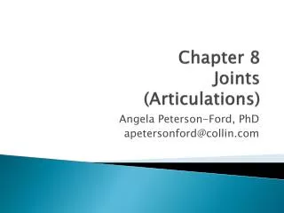
Chapter 9: Joints
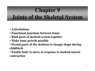
Chapter 9: Joints. Anatomy 32. I. Articulations: bones are rigid structures but become moveable at the joint or articulations (Greek- arthro). Joints may occur as bone to bone, bone to cartilage, or teeth to bone.

Chapter 9: Joints
E N D
Presentation Transcript
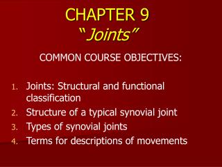
Chapter 9: Joints Anatomy 32
I. Articulations: bones are rigid structures but become moveable at the joint or articulations (Greek- arthro). Joints may occur as bone to bone, bone to cartilage, or teeth to bone. A. Classification of Joints- when classified by function the focus is placed one the amount of movement. When classified by structure the focus is on the tissue type that makes the joint. See table 9.1 pg 202for structural and functional (mobility) characteristics. 1. synarthrosis- immovable 2. amphiarthrosis- slightly moveable 3. diarthroses-freely moveable
B. Fribrous joints- These are immobile or minutely mobile. Examples are the joints of the cranium and the teeth. They are lined with fibrous tissues (dense regular tissue). 1. Sutures- immoveable joints formed by skull bones and connected by fibrous tissue.
2. Syndesmosis- slightly moveable but no true movement, formed by a ligament band of fibrous tissue
3. Gomphoses-peg in socket as in the tooth and gum containing a small ligament
C. Cartilaginous joints-articulating bones are united by cartilage, no joint cavity, not highly moveable. 1. Synchondroses- hyaline cartilage that unites bones as in the ribs and sternum
2. Symphyses- fibrocartilage that unites bones as in the pubic symphysis and intervertebral bones.
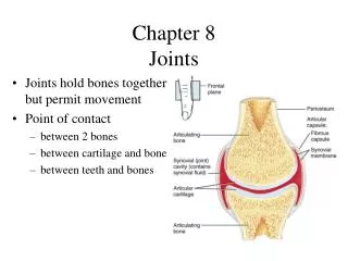
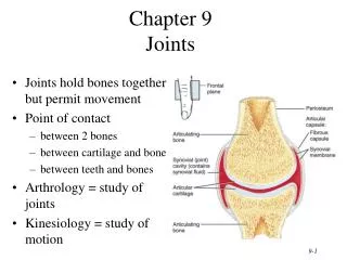
D. Synovial joints-most moveable, has fluid filled cavity with synovial fluid, called diarthrosis. 1.Basic Features-hyaline cartilage lines the ends of bones, a cavity filled with fluid called synovial fluid is contained by a fibrous capsule. It that has an outer layer of dense irregular tissue and joins the periosteum and an inner layer of synovial membrane (makes synovial fluid). The synovial fluid is viscous liquid that lubricates the joint. Ligaments reinforce the joint and nerves protect it from over stretching. Some joints also have a meniscus (articular discs) such as the knee joint.
Synovial Joint with articulate disc (meniscus) in the middle
2. Function (mobility)- these joints are highly lubricated to facilitate motion, they routinely experience compression and the fluid moves to accommodate the pressure. 3. Joint stability- some joints have articulating surfaces that stabilize the joints such as the elbow and hip joint. Ligaments strengthen the joint and prevent it from moving incorrectly, they are not as flexiable and capable of reshaping. 4. Joint classification- the following classifications are based on joint shape. a. plane joints- flat articulating surfaces that allow for short gliding movements as in the wrist bones (intercarpals), ankle bones (intertarsals), and vertebral bones, no rotation around an axis (non-axial).
b. hinge joints- movement is along one plane (uniaxial) such a hinged door. Examples are the elbow and knee.
c. pivot joints- rounded end of one bone fits into the other, also uniaxial, examples are the radius and ulna and atlas and dens.
d. condyloid joints-(knuckle like) oval like shape of one bone fits into an oval bowl like shape of the other. It does not rotate around its axis but can move sided to side and back and forth, it is biaxial.
e. saddle joints- the two ends fit into one another as a person sitting in a saddle does. They are biaxial such as the thumb joint (first carpometacarpal joint)
f. ball and socket joints- spherical end fits into round socket, allows movement in multiple planes of axis (triaxial) such as the hip and shoulder.
5. Selected synovial joint a. temporomandibular joint- this is a synovial joint that has an articular capsule, hinge like movement, and anterior movement. Because it is so shallow it can be easily dislocated.
b. shoulder joint- less stable but more moveable, the glenoid fossa is lined with fibrocartilage called the genoid labrum, ligaments support limb weight and muscle tendons contribute to stability. This joint has a rotator cuff formed by tendons. It also has bursa and tendon sheaths.
c. elbow joint- stable hinge joint, ligaments stabilize and prevent lateral/medial movements.
Lateral View Medial View
d. hip joint- stable ball-and-socket joint, wide range of motion but less than shoulder joint due to more depth. Also has a rim of fibrocartilage called acetabular labrum that prevents the femur from slipping. Capsular ligaments provide stability.
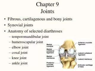
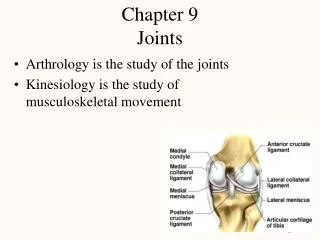
Knee Joint Information e. knee joint- largest and most complex, it includes the femur, tibia, and patella. It contains: 1) several bursae, 2) lateral and medial menisci (fibrocartilage rings) that guide the condyles. 3) three anterior ligaments originating from patella to tibia: patellar ligament, medial and lateral ligament 4) fibular and tibial collateral ligaments support the joint capsule 5) anterior and posterior cruciate- attaches across joint internally 6) ligaments from fibula to femur anterior or posterior
Anterior view of the knee. A lateral blow can cause the femur and tibia to separate tearing several ligaments that are attached to each other. Pg. 234
E. Joint disorders 1. Sprain-stretch or torn ligaments 2. Dislocation (luxation)- bones are forced out of alignment and must be reduced (returned to original location). Subluxation is a partial dislocation. 3. Torn cartilage- when it is subjected to high compression and tension, example: meniscus tearing. It does not heal itself thus broken fragments are removed by arthroscopic surgery. 4. bursitis/tendonitis- swelling of the bursa or tendon sheaths 5. arthritis- inflammation or degeneration of the joints. a. osteoarthiritis- related to aging, the joints wear out. Use of joint causes enzyme that breakdown the cartilage to breakdown, occurs more often in misaligned joints may form bone spurs. It happens often in non-synovial joints.
b. rheumatoid arthiritis- it is an autoimmune disease in which the immune system attacks the cartilage causing inflammation and muscle weakness. It affect women more than man and joints bilaterally and simultaneously. c. gouty arthiritis- uric acid accumulates in joints causing inflammation at the joints. It affects men more than women and if untreated can lead to fused bones. d. Lyme disease-bacteria transmitted by tick bites causes joint inflammation and many other symptoms. It is difficult to diagnose and treat. F. Joints throughout life Synovial joints originate from mesenchyme, joints are modified after use by movement and use- more active joints are more massive. Epiphyseal plates are vulnerable during youth. With aging the joint become arthritic although this can be delayed with exercise. Good luck preparing for the test!!!!!
Movement of synovial joints- Angular movements- increase or decrease the joint angle and bring limb towards or away 1. Flexion- reduces joint angle (bending at the joint) brings bones closer, usually in the sagittal plane 2. Extension- increases joint angle (straightening joint) brings bones further apart 3. Hyperextension- increasing joint angle beyond its normal range 4. Abduction- moving the joint such that limbs are moved away from the body 5. Adduction- moving the joint such that limbs are moved toward the body 6. Circumduction- a combination of flexion, extension, abduction, and adduction. (making a circle) 7. Rotation-movement of bone along its own long axis only three areas can do this: atlas and axis, shoulder joint, hip joint. Medial rotation- movement towards the median . Lateral rotation- movement away from the median 8. Supination- lateral radial rotation to turn palm anteriorly 9. Pronation-medial radial rotation to turn palm posteriorly 10. Dorsiflexion-lifting foot to superior side of foot approaches shin 11. Plantar flexion-depressing the foot (pointing toes) 12. Inversion-turn sole of foot medially 13. Eversion-turn sole of foot laterally 14. Protraction and Retraction- non angular movement, forward= protaction back=retraction 15. Elevation- moving body part superiorly 16. Depression- moving body part inferiorly
1. Flexion- reduces joint angle (bending at the joint) brings bones closer, usually in the sagittal plane 2. Extension- increases joint angle (straightening joint) brings bones further apart 3. Hyperextension- increasing joint angle beyond its normal range
1. Flexion- reduces joint angle (bending at the joint) brings bones closer, usually in the sagittal plane 2. Extension- increases joint angle (straightening joint) brings bones further apart 3. Hyperextension- increasing joint angle beyond its normal range
1. Flexion- reduces joint angle (bending at the joint) brings bones closer, usually in the sagittal plane 2. Extension- increases joint angle (straightening joint) brings bones further apart 3. Hyperextension- increasing joint angle beyond its normal range
4. Abduction- moving the joint such that limbs are moved away from the body 5. Adduction- moving the joint such that limbs are moved toward the body 6. Circumduction- a combination of flexion, extension, abduction, and adduction. (making a circle) 7. Rotation-movement of bone along its own long axis only three areas can do this: atlas and axis, shoulder joint, hip joint. Medial rotation- movement towards the median . Lateral rotation- movement away from the median
8. Supination- lateral radial rotation to turn palm anteriorly 9. Pronation-medial radial rotation to turn palm posteriorly 10. Dorsiflexion-lifting foot to superior side of foot approaches shin 11. Plantar flexion-depressing the foot (pointing toes)
12. Inversion-turn sole of foot medially 13. Eversion-turn sole of foot laterally 14. Protraction and Retraction- non angular movement, forward= protaction back=retraction
15. Elevation- moving body part superiorly 16. Depression- moving body part inferiorly