Cells: the basic structural & functional units of living things
420 likes | 579 Vues
Cells: the basic structural & functional units of living things plasma membrane : flexible outer surface of cell; selective barrier that regulates flow of materials into & out of cell – maintains internal environment cytoplasm : all cellular contents between plasma membrane & nucleus

Cells: the basic structural & functional units of living things
E N D
Presentation Transcript
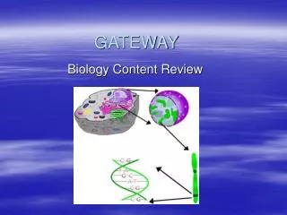
Cells: the basic structural & functional units of living things • plasma membrane: flexible outer surface of cell; selective barrier that regulates flow of materials into & out of cell – maintains internal environment • cytoplasm: all cellular contents between plasma membrane & nucleus • contains organelles: small, membrane-bounded bodies with a specific structure & function (e.g.: mitochondria, chloroplasts, lysosomes) in cytosol (semifluid medium between nucleus and plasma membrane) • nucleus: large organelle that stores DNA in the form of chromosomes containing genes
Cell membrane: outer boundary of cells • phospholipid bilayer: semipermeable and selectively permeable • functions in regulation of passage of molecules into and out of the cell • membrane components: • phospholipids: create bilayer • have polar & nonpolar parts • glycolipids: protective function, and cell identity (specific for cell type) • cholesterol: bulky; controls (reduces) permeability • proteins: also glycoproteins; can be transmembrane (spans the entire membrane) or embedded in either the cytoplasmic or extracellular side of the membrane • glycoproteins (and glycolipids) function in cell-cell recognition (cell fingerprint); important in transplantation • Plasma Membrane is semipermeable and selectively permeable: some molecules may pass through freely (e.g.: water); others must be assisted across
Types of Membrane Proteins: • Channel Proteins: create transient hydrophilic channel for small molecules & ions to flow into & out of cell • Carrier Proteins: selectively interact with small molecules or ions to assist them across the membrane • Cell Recognition Protein: Cell Identity; individual-specific groups of proteins on extracellular side of membrane (e.g.: MHC/HLA (Human Leukocyte Antigen) – important to match with donor to avoid rejection of transplanted organ or tissue) • Receptor Protein: Interacts with specific molecule to transmit some type of signal or communication (electrical, chemical or contact) between cells (e.g.: hormone receptors) • Enzymatic Protein: Catalyzes (speeds up) some specific reaction which results in a cellular response • Cell Adhesion Molecules (CAMs): guide interactions between cells
Cytosol: consists of cytoplasm (the fluid within the cell outside the nucleus) & organelles • Endoplasmic Reticulum: (ER) • Rough ER: associated with ribosomes; proteins translated on ribosomes associated with the rough ER will be transported and/or secreted outside cell • begins processing & modification of these proteins • Smooth ER: synthesizes phospholipids in all cells; various other cell type-specific functions • synthesizes steroid hormones in testes, and detoxifies drugs in liver cells • Ribosomes: site of protein synthesis in the cell • free in cytoplasm (polyribosomes) or associated with rough endoplasmic reticulum • 2 subunits (large & small); mRNA is threaded through subunits during translation (protein synthesis) • Golgi apparatus: completes modification of proteins from rough ER (proteins transported to Golgi in vesicles) • modification of proteins & lipids (addition of carbohydrate chains (glycosylation)) • also transports organic molecules in vesicles; some become lysosomes
Mitochondria: produces energy • site of cellular respiration (ATP production from carbohydrates) • also have folded membrane system (folds are cristae, inner fluid-filled space is the matrix) • extensive membrane systems are important in both chloroplasts and mitochondria for ATP production
Lysosomes: vesicles with digestive enzymes to break down macromolecules & cell debris • loss of some or all lysosome function in inherited disorders (Tay-Sachs disease) may lead to accumulation of unwanted molecules (& related toxicity) • Peroxisomes are vesicles that contain enzymes for oxidizing certain organic molecules with the release of hydrogen peroxide (toxic, but breaks down into water & oxygen)
Cytoskeleton: composed of microfilaments, microtubules, & intermediate filaments • functions in maintaining shape of cell and movement of subcellular structures • microfilaments: thinnest elements of cytoskeleton; help generate movement & provide mechanical support • actin filaments combine with myosin in muscle cells to enable muscle movement • microtubules: composed of tubulin dimers coiled into tubelike structures • concentrated & arranged as rings of nine doublets or triplets in centrioles, cilia, and flagella • microtubules involved in movement
Centrosome: located near nucleus; consists of centrioles & pericentriolar material • centrioles: cylindrical structures composed of 9 clusters of three microtubules (triplets) arranged in circular pattern • pericentriolar material consists of hundreds of tubulin complexes • involved in organization of spindle fibers for chromosome movement during mitosis
Cilia and Flagella: composed of microtubules (9 + 2 pattern); used in movement • Cilia present in some unicellular protists (Paramecium) and cells of respiratory tract in animals • Flagella present in some unicellular protists (Euglena) and sperm cells
Vesicles (vacuoles): membrane-bounded organelles for transport or storage • formed by cell membrane, ER or Golgi apparatus
Nucleus: stores genetic information in all eukaryotic cells • DNA is organized into distinct chromosomes • Chromosomes are packaged with proteins to form chromatin • dark regions within the nucleus are nucleoli (1 or more per cell) • within each nucleolus, ribosomal RNA is produced and joins with ribosomal proteins to form ribosomes • the nucleus is bounded by a porous membrane, the nuclear envelope, which regulates passage of molecules into & out of the nucleus
Plasma membrane transport: • Diffusion: movement of molecules from a region of higher concentration to a region of lower concentration (down concentration gradient) • evenly distributes molecules in water (equilibrium) • lipid soluble molecules, gases (oxygen, carbon dioxide) and water can diffuse across the plasma membrane
Facilitated Diffusion: passage of small molecules (glucose, amino acids) across the plasma membrane even though they may not be lipid-soluble • a carrier protein assists movement of molecules down concentration gradient • no energy is required
Osmosis: diffusion of water across a differentially permeable membrane (plasma membrane) • important in water retention
Tonicity: the strength (solute concentration) of a solution in relation to osmosis • in cells, the solute concentration of a solution with respect to that solute concentration inside the cell • isotonic (iso-osmotic) solution: the net solute concentration of the solution equals that inside the cell • hypotonic (hypo-osmotic) solution: the net solute concentration of the solution is less that inside the cell; animal cells swell (& eventually will burst – hemolysis) • hypertonic (hyper-osmotic) solution: the net solute concentration of the solution is greater that inside the cell; animal cells shrink – crenation
Filtration: a pressure gradient pushes solute-containing fluid (filtrate) from area of high pressure to area of low pressure • forces water & solutes through membrane or capillary wall by hydrostatic pressure