BASICS OF PACEMAKER
BASICS OF PACEMAKER. DN. HISTORY. 1958 – Senning and Elmqvist Asynchronous (VVI) pacemaker implanted by thoracotomy and functioned for 3 hours Arne Larsson First pacemaker patient


BASICS OF PACEMAKER
E N D
Presentation Transcript
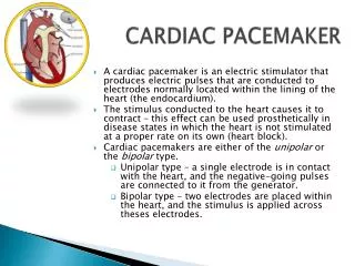
HISTORY • 1958 –Senning and Elmqvist • Asynchronous (VVI) pacemaker implanted by thoracotomy and functioned for 3 hours • Arne Larsson • First pacemaker patient • Used 23 pulse generators and 5 electrode systems • Died 2001 at age 86 of cancer • 1960 – First atrial triggered pacemaker • 1964 – First on demand pacemaker (DVI) • 1977 – First atrial and ventricular demand pacing (DDD) • 1981 – Rate responsive pacing by QT interval, respiration, and movement • 1994 – Cardiac resynchronization pacing
What is a Pacemaker? A Pacemaker System consists of a Pulse Generator plus Lead (s)
Implantable Pacemaker Systems Contain the Following Components: • Pulse generator- power source or battery • Leads • Cathode (negative electrode) • Anode (positive electrode) • Body tissue Lead IPG Anode Cathode S
The Pulse Generator • Contains a battery that provides the energy for sending electrical impulses to the heart • Houses the circuitry that controls pacemaker operations Circuitry Battery
Anatomy of a Pacemaker Resistors Atrial connector Connector Ventricular connector Defibrillation protection Output capacitors Hybrid Clock Reed (Magnet) switch Telemetry antenna Battery
General Characteristics of Pacemaker Batteries • Hermeticity, as defined by the pacing industry, is an extremely low rate of helium gas leakage from the sealed pacemaker container • low rate of self-discharge • lithium iodine -a long shelf life and high energy density • DDD drains a battery more rapidly
Power source • Longevity in single chamber pacemaker is 7 to 12 years. • For dual chamber longevity is 6 to 10 years. • Most pacemakers generate 2.8 v in the beginning of life which becomes 2.1 to 2.4 v towards end of life.
Leads • Deliver electrical impulses from the pulse generator to the heart • Sense cardiac depolarisation Lead
Position within the heart Endocardial or transvenous leads Epicardial leads Fixation mechanism Active/Screw-in Passive/Tined Shape Straight J-shaped used in the atrium Polarity Unipolar Bipolar Insulator Silicone Polyurethane Lead Characterization
Lead components • Conductor • Connector Pin • Insulation • Electrode
Passive fixation • The tines become lodged in the trabeculae
Active Fixation • The helix (or screw) extends into the endocardial tissue • Allows for lead positioning anywhere in the heart’s chamber
Myocardial and Epicardial Leads • Leads applied directly to the heart • Fixation mechanisms include: • Epicardial stab-in • Myocardial screw-in • Suture-on
Cathode:-An electrode that is in contact with the heart • Negatively charged • Anode:-receives the electrical impulse after depolarization of cardiac tissue • Positively charged when electrical current is flowing Anode Cathode
A Unipolar Pacing System Contains a lead with an electrode in the heart • Flows through the tip electrode (cathode) • Stimulates the heart • Returns through body fluid and tissue to the PG (anode) + Anode - Cathode
A Bipolar Pacing System Contains a lead with 2 electrodes in the heart • Flows through the tip electrode located at the end of the lead wire • Stimulates the heart • Returns to the ring electrode above the lead tip Anode Cathode
One electrode on the tip & one conductor coil • Conductor coil may consist of multiple strands - (multifilar leads) Unipolar leads • Unipolar leads have a smaller diameter than bipolar leads • Unipolar leads exhibit larger pacing artifacts on the surface ECG
Bipolar leads • Circuit is tip electrode to ring electrode • Two conductor coils (one inside the other) • Inner layer of insulation • Bipolar leads are typically thicker than unipolar leads • Bipolar leads are less susceptible to oversensingnoncardiac signals (myopotentials and EMI) Coaxial Lead Design
Electrodes • Leads have 1/> electrically active surfaces referred to as the electrodes • Deliver an electrical stimulus, detect intrinsic cardiac electrical activity, or both • Electrode performance can be affected by • Materials • Polarization • Impedance • Pacing thresholds • Steroids
Electrode Materials • The ideal material for an electrode • Porous (allows tissue ingrowth) • Should not corrode or degrade • Small in size but have large surface area • Common materials • Platinum and alloys (titanium-coated platinum iridium) • Vitreous carbon (pyrolytic carbon) • Stainless steel alloys such as Elgiloy
Voltage • Voltage is the force that causes electrons to move through a circuit • In a pacing system, voltage is: • Measured in volts • Represented by the letter “V” • Provided by the pacemaker battery • Referred to as amplitude
Current • The flow of electrons in a completed circuit • In a pacing system, current is: • Measured in mA (milliamps) • Represented by the letter “I” • Determined by the amount of electrons that move through a circuit
Constant-Voltage and Constant-Current Pacing • Most permanent pacemakers are constant-voltage pacemakers • Voltage and Current Threshold • Voltage threshold is the most commonly used measurement of pacing threshold
Pacing Thresholds • Defined as the minimum amount of electrical energy required to consistently cause a cardiac depolarization • “Consistently” refers to at least ‘5’ consecutive beats • Low thresholds require less battery energy Capture Non-Capture
2.0 1.5 1.0 .50 .25 The Strength-Duration Curve • The strength-duration curve illustrates the relationship of amplitude and pulse width • Values on or above the curve will result in capture Stimulation Threshold (Volts) Capture 0.5 1.0 1.5 Duration Pulse Width (ms)
Rheobase- (the lowest point on the curve) by definition is the lowest voltage that results in myocardial depolarization at infinitely long pulse duration • Chronaxie(pulse duration time ) by definition, the chronaxie is the threshold pulse duration at twice the rheobase voltage
Lessons from SDC • The ideal pulse duration should be greater than the chronaxie time • Cannot overcome high threshold exit block by increasing the pulse duration, If the voltage output remains less than the rheobase • Energy (μJ) = Voltage (V) × Current (mA) × Pulse Duration (PD in ms). • Charge (μC) = Current (mA) × Pulse Duration (ms).
At very low pulse width thresholds, the charge is low, but the energy requirements are high because of elevated current and voltage stimulation thresholds. • At pulse durations of 0.4–0.6 ms, all threshold parameters - ideal • At high pulse durations, the voltage and current requirements may be low, but the energy and charge values are unacceptable
-Safety margins -When a threshold is determined by decrementing the pulse width at a fixed voltage • At a given voltage where the pulse width value is < .30 ms: Tripling the pulse width will provide a two-time voltage safety margin. • Daily fluctuations in threshold that can occur due to eating, sleeping, exercise, or other factors - a/c pacing system - higher safety margin, due to the lead maturation process- occur within the first 6-8 weeks following implant.
Changes in stimulation threshold (voltage or current) following implantation of a standard nonsteroid-eluting electrode
Impedance • The opposition to current flow • In a pacing system, impedance is • Measured in ohms • Represented by the letter “R” (W for numerical values) • The measurement of the sum of all resistance to the flow of current Resistance is a term used to refer to simple electric circuits without capacitors and with constant voltage and current Impedance is a term used to describe more complex circuits with capacitors and with varying voltage and current
Impedance • Pacing lead impedance typically stated in broad ranges, i.e. 300 to 1500 Ω • Factors that can influence impedance • Resistance of the conductor coils • Tissue between anode and cathode • The electrode/myocardial interface • Size of the electrode’s surface area • Size and shape of the tip electrode
V I R Ohm’s Law is a Fundamental Principle of Pacing That: • Describes the relationship between voltage, current, and resistance V = I X R I = V / R R = V / I x
Impedance and Electrodes • Large electrode tip • Threshold ↑ • Impedance ↓ • Polarization ↓ • Small electrode tip • Threshold ↓ • Impedance ↑ • Polarization ↑
Polarization • After an output pulse, positively charged particles gather near the electrode. • The amount of positive charge is • Directly proportional to pulse duration • Inversely proportional to the functional electrode size (i.e. smaller electrodes offer higher polarization) Polarization effect can represent 30–40% of the total pacing impedance As high as 70% for smooth surface, small surface area electrodes
Within the electrode, current flow is due to movement of electrons (e−). At the electrode–tissue interface, the current flow becomes ionic & (-) vely charged ions (Cl−, OH−) flow into the tissues toward the anode leaving behind oppositely charged particles attracted by the emerging electrons. It is this capacitance effect at the electrode tissue interface, that is the basis of polarization
Lead Maturation Process • Fibrotic “capsule” develops around the electrode following lead implantation • 3 phases • A/c phase, where thresholds immediately following implant are low • Peaking phase- thresholds rise and reach their highest point(1wk) ,followed by a ↓ in the threshold over the next 6 to 8 wks as the tissue reaction subsides • C/c phase- thresholds at a level higher than that at implantation but less than the peak threshold • Trauma to cells surrounding the electrode→ edema and subsequent development of a fibrotic capsule. • Inexcitable capsule ↓ the current at the electrode interface, requiring more energy to capture the heart.
5 4 Smooth Metal Electrode 3 Volts Textured Metal Electrode 2 1 Steroid-Eluting Electrode 0 1 2 4 5 7 8 9 10 11 12 Implant Time (Weeks) Lead Maturation Process • Effect of Steroid on Stimulation Thresholds 0 3 6 Pulse Width = 0.5 msec
Sensing • Sensing is the ability of the pacemaker to detect an intrinsic depolarization • Pacemakers sense cardiac depolarization by measuring changes in electrical potential of myocardial cells between the anode and cathode
An Electrogram (EGM) is the Recording of Cardiac Waveforms Taken From Within the Heart • Intrinsic deflection on an EGM occurs when a depolarization wave passes directly under the electrodes • Two characteristics of the EGM are: • Signal amplitude(mv) • Slew rate(v/sec)
Intrinsic R wave Amplitude • Typical intrinsic R wave amplitude measured from pacing leads in the Right Ventricle are more than 5 mV in amplitude Intrinsic R wave in EGM Amplitude The Intrinsic R wave amplitude is usually much greater than the T wave amplitude
Slew Rate of the EGM Signal Measures the Change in Voltage with Respect to the Change in Time • The longer the signal takes to move from peak to peak: • The lower the slew rate • The flatter the signal • Higher slew rates translate to greater sensing • Measured in volts per second Change in voltage Slew rate= Time duration of voltage change Voltage Slope Time Slew rate measurements at implant should exceed .5 volts per second for P waves; .75 volts per second for R wave measurements