FUNCTIONAL MRI in MOTOR IMAGERY
340 likes | 693 Vues
FUNCTIONAL MRI in MOTOR IMAGERY. JEFFREY S. ROSS, M.D. NEURORADIOLOGY CLEVELAND CLINIC FOUNDATION ROSSJ1@CCF.ORG. FUNCTIONAL MRI. BRAIN IS UNIQUE Strong relation between location and function MULTIPLE TECHNIQUES TO EVALUATE FUNCTION Clinical cases (strokes, tumors, epilepsy, etc.)

FUNCTIONAL MRI in MOTOR IMAGERY
E N D
Presentation Transcript
FUNCTIONAL MRIin MOTOR IMAGERY JEFFREY S. ROSS, M.D. NEURORADIOLOGY CLEVELAND CLINIC FOUNDATION ROSSJ1@CCF.ORG
FUNCTIONAL MRI • BRAIN IS UNIQUE • Strong relation between location and function • MULTIPLE TECHNIQUES TO EVALUATE FUNCTION • Clinical cases (strokes, tumors, epilepsy, etc.) • Animal lesion studies • EEG, MEG, etc. • Direct intraoperative electrical stimulation • Functional imaging (PET, FMRI)
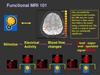
BASICS Change in local brain activity Change in local brain metabolism Change in local hemodynamics
BASICS • IMAGE DURING 2 BEHAVIORAL STATES • Control state • Activation state • “DIFFERENCE” IMAGE SHOWS “ACTIVATED” BRAIN • Brain activated depends critically on design of task • Tasks carefully designed to isolate function of interest • BLOOD FLOW USED AS ENDOGENOUS CONTRAST
BOLD FMRI basics • BOLD: Blood Oxygenation Level Dependent • Signal changes based on blood O2 level • Oxyhemoglobin (HbO2) diamagnetic • Deoxyhemoglobin (Hb) weakly paramagnetic • Paramagnetic substance • Causes local magnetic field heterogeneity • Shortens T2, T2* relaxation times • Lowers signal in tissue
BOLD effect 100% O2 Room air Rat brain at 9 tesla (Ogawa et al MRM 14:68, 1990)
BOLD EFFECT • BLOOD O2 LEVEL • Blood flow • Tissue metabolism • NEURAL ACTIVATION • Increase in local blood flow greater than • Increase in local O2 extraction • Increased capillary, venous HbO2 • More HbO2 causes increased signal • SMALL • DELAYED WITH RESPECT TO STIMULUS ONSET • LASTS A FEW SECONDS • FIELD STRENGTH DEPENDENT
BOLD EFFECT RESTING ACTIVATED CBV O2 SAT CBF ARTERY CAP VEIN
fMRI signal • SIGNAL CHANGES OF 2-5% • STRONGLY FIELD DEPENDENT • LAG TIME FOR RESPONSE, AT LEAST 300 msec UP TO 4s off on off on off 0
PROBLEMS • IMAGE NOISE • MOTION SENSITIVE • Head motion under 0.5 mm can ruin data • SILENT REGIONS • MAKE NOTHING OF WHAT YOU DON’T SEE • ACTIVATED REGIONS • ACTUAL CONTRIBUTION TO COGNITIVE TASK? • ANATOMICAL ACCURACY • LOCALIZATION OF EXTENT OF ACTIVATION • ASSUME ACTIVATION TAKES FORM OF DISCRETE LOCAL INCREASE IN FLOW
PROBLEMS • PARADIGM DESIGN • ASSUME BRAIN IN STABLE COGNITIVE STATE DURING MEASUREMENT • HOW OFTEN REPEATED • HOW EASY/HARD TO PERFORM • NATURAL OR TIGHTLY CONTROLLED • COGNITIVE DEMANDS • EMOTIONS • MACROSCOPIC LANDMARKS • SEVERAL “SPACES” – TALAIRACH • PRECISE TEMPORAL RELATIONSHIP
TONGUE LIP HAND ELBOW SHOULDER HIP KNEE ANKLE Mapping the Sensori-motor Homunculus
MOTOR IMAGERY -Introduction • Mental imagery involves rehearsing or practicing a task in the mind with no physical movement • The technique is commonly utilized, and widely advocated • Physical foundation of imagery not well characterized for fast, complex, automatic motor movements such as the golf swing
It's a great image for any golfer faced with a pressure shot. Replay an old "tape" from your memory bank, thinking of a past success rather than calling up a negative memory. You'll then have an excellent chance of duplicating the successful shot. LEADBETTER’S IMAGES GOLF DIGEST, MARCH 2001
Introduction • This study evaluated motor imagery of the golf swing, of golfers of various handicaps, using fMRI to: • 1) Assess whether areas of brain activation could be defined by this technique and; • 2) To define any association between activated brain areas and golf skill.
Materials and Methods • Six golfers of various handicap levels (0,5,7,10,11,13) with evaluated with functional MRI during a control condition and during mental imagery of their golf swing. • Five of the six subjects were right handed, and play golf right handed. • The subject with a 5 handicap (hcp) was left handed, but plays golf right handed. • 6 males, 24-50 years of age, average age 39 years.
Materials and Methods • 1st person perspective, as they would on a practice tee, with each swing mentally occurring every 1.5-2 seconds. No preshot routine. • Two control conditions were evaluated, “rest” and “wall”
PARADIGMS • REST • subjects were told to project themselves into a restful state, such as sitting quietly on a beach, taking care not to move mentally (or physically) during the study • WALL • were told to imagine leaning against a wall with their hands outstretched and pushing against it
Materials and Methods • Functional studies were performed on either a 1.5T whole body (Symphony, Siemens, Erlangen) or 3T head only (Allegra, Siemens, Erlangen) MRI systems. • On the 1.5T system, the body coil was used to transmit, with a receive-only head coil collecting the data. • A transmit/receive head coil was used to acquire the 3T functional studies. • Functional images : 2D multi-slice gradient echo EPI acquisition. Sixteen slice locations were collected every 3-4 sec with fat saturation.
RESULTS • Vermis, SMA, cerebellum and motor regions generally showed the greatest activation • Little activation seen in cingulate gyrus, right temporal lobe, deep gray matter, and brainstem • Wall vs. golf paradigm showed generally diminished activation across all regions compared to the Rest vs. golf • Decreased activation most extensively with the better players
RESULTS • Assuming a limit of 2% activation, 95% confidence intervals were generated. This showed with a 95% confidence that the following areas were activated greater than 2% • 13 handicap: Brainstem, Left motor, Left sensory, Left SMA, Left temporal, Right cerebellum, Right SMA, Vermis • 7 handicap: Left SMA • 5 handicap: no areas • 0 handicap: no areas
Percent area activation, 13 handicap averaged five series of “rest vs. golf”
REST VS GOLF 13 HANDICAP
CONCLUSIONS • Demonstrated the feasibility of defining areas of brain activation during imagery of a complex, coordinated motor task • Suggests increased activation with increasing handicap
CONCLUSIONS • Generally good agreement of golf swing imagery with brain activation areas defined in the literature including: • Primary motor control (motor cortex) • Imagery (parietal cortex) • Execution areas (premotor cortex of frontal lobe, lateral cerebellum, basal ganglia, vermis and medial cerebellar hemispheres) • Action planning areas (frontal and parietal cortex, SMA, lateral cerebellum) • Error detection (cingulate, cerebellum)
CONCLUSIONS • Increased activation with high handicap players could potentially relate to two effects: • 1) that increased activation reflects a failure to learn and become highly automatic or • 2) that increased activation is essentially pathologic, and related to a loss of automaticity with compensatory increased brain activity. • Development of automaticity is relative and can be dynamic and reversible • Classic example of this pathology is writer’s cramp (focal dystonia) where the severe functional disturbances can be explained in terms of a loss of automaticity and an increased need for controlled processing
Ross JS et al. The Minds Eye: Functional imaging of golf motor imagery. American Journal of Neuroradiology, 24:1036-1044, June/July 2003. QUESTIONS? rossj1@ccf.org