Protein-Nucleic Acid Dynamics
400 likes | 587 Vues
Protein-Nucleic Acid Dynamics. Ashok Kolaskar Vice Chancellor University of Pune Pune India. Molecular Dynamics: Introduction. Biomolecules are polymers of basic building blocks Proteins Amino Acids Nucleic acids Nucleotides Carbohydrates Sugars.

Protein-Nucleic Acid Dynamics
E N D
Presentation Transcript
Protein-Nucleic Acid Dynamics Ashok Kolaskar Vice Chancellor University of Pune Pune India
Molecular Dynamics: Introduction Biomolecules are polymers of basic building blocks Proteins Amino Acids Nucleic acids Nucleotides Carbohydrates Sugars
Molecular Dynamics: Introduction • At physiological conditions, the biomolecules undergo several movements and changes • The time-scales of the motions are diverse, ranging from few femtoseconds to few seconds • These motions are crucial for the function of the biomolecules
Molecular Dynamics: Introduction Newton’s second law of motion
Molecular Dynamics: Introduction We need to know The motion of the atoms in a molecule, x(t) and therefore, the potential energy, V(x)
Molecular Dynamics: Introduction How do we describe the potential energy V(x) for a molecule? Potential Energy includes terms for Bond stretching Angle Bending Torsional rotation Improper dihedrals
Molecular Dynamics: Introduction Potential energy includes terms for (contd.) Electrostatic Interactions van der Waals Interactions
Molecular Dynamics: Introduction Equation for covalent terms in P.E.
Molecular Dynamics: Introduction Equation for non-bonded terms in P.E.
Molecular Dynamics: Introduction • Each of these interactions exerts a force onto a given atom of the molecule • The total resulting force on each atom is calculated using the PE function Knowing the force on an atom, its movement due to the force is then calculated:
Molecular Dynamics: Introduction To do this, we should know at given time t, • initial position of the atom x1 • its velocity v1 = dx1/dt • and the acceleration a1 = d2x1/dt2 = m-1F(x1)
Molecular Dynamics: Introduction The position x2 , of the atom after time interval t would be, and the velocity v2 would be,
Molecular Dynamics: Introduction In general, given the values x1, v1 and the potential energy V(x), the molecular trajectoryx(t)can be calculated, using,
Generalizing these ideas, the trajectories for all the atoms of a molecule can be calculated.
The Necessary Ingredients • Description of the structure: atoms and connectivity • Initial structure: geometry of the system • Potential Energy Function: force field • AMBER • CVFF • CFF95 • Universal
Protein-specific Applications of MD • Calculation of thermodynamic properties such as internal energy, free energy • Studying the protein folding / unfolding process • Studying conformational properties and transitions due to environmental conditions • Studying conformational distributions in molecular system.
A typical MD simulation protocol • Initial random structure generation • Initial energy minimization • Equilibration • Dynamics run – with capture of conformations at regular intervals • Energy minimization of each captured conformation
Essential Parameters for MD (to be set by user) • Temperature • Pressure • Time step • Dielectric constant • Force field • Durations of equilibration and MD run • pH effect (addition of ions)
WHAT IS AMBER? • AMBER (Assisted Model Building with Energy Refinement). • Allows users to carry out molecular dynamics simulations • Updated forcefield for proteins and nucleic acids • Parallelized dynamics codes • Ewald sum periodicity • New graphical and text-based tools for building molecules • Powerful tools for NMR spectral simulations • New dynamics and free energy program
WHY AMBER? • Most widely used program: approximately 5000 users world over. • Over 1000 research papers have been published using AMBER. • Program available at a nominal price for academic users. • Complete source code available with the package. • Available for most machine configurations. • Developed by Prof.Peter Kollman at the University of California San Francisco: An authority in the area of molecular simulations.
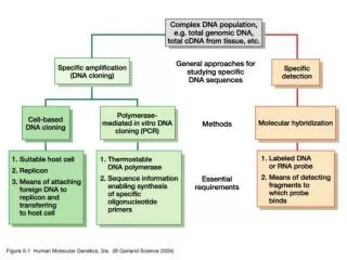
BASIC INFORMATION FLOW IN AMBER database seq pdb forcefield prep link edit parm nmode constraints Nmanal, lmanal Sander, Gibbs, spasms carnal anal mdanal
CASE STUDY • Type II restriction endonucleases recognize DNA sequences of 4 to 8 base pairs in length and require Mg2+ to hydrolyse DNA. • The recognition of DNA sequences by endonucleases is still an open question. • PvuII endonuclease, recognizes the sequence 5’-CAGCTG-3’ and cleaves between the central G and C bases in both strands. • Though crystal structure of the PvuII-DNA complex have been reported, very little is known about the steps involved in the recognition of the cleavage site by the PvuII enzyme. • Molecular dynamics (MD) simulation is a powerful computational approach to study the macromolecular structure and motions.
CASE STUDY: METHODS (MD Simulations) • Simulations were carried out on the sequence • 5’-TGACCAGCTGGTC-3’ • Rectangular box (60 X 48 X 54 Å3) containing 24 Na+, using PBC • SHAKE algorithm • Integration time step of 1 fs • 283 K with Berendsen coupling • Particle Mesh Ewald (PME) method • 9.0 Å cutoff was applied to the Lennard-Jones interaction term. • Equilibration was performed by slowly raising the temperature from 100 to 283 K. Production run was initiated for 1.288 ns and the structures were saved at intervals of one picosecond. • The trajectory files were imaged using the RDPARM program and viewed and analysed using the MOIL-VIEW and CURVES packages respectively.
RESULTS Particle Mesh Ewald simulations of PvuII substrate • The simulations carried out using PME method, points out that the initial straight B-helix conformation bends significantly as the simulation progresses. The DNA molecule bends maximally by 18% and 22% at 616 ps and 1243 ps respectively. The base pair rise (h) between G7:C7’ and C8:G6’ observed in this simulation, shows large fluctuations around the normal value. • The average roll value is seen to increase with simulation time and this indicates bending of the DNA molecule. • The offset values, for each base pair showed that the maximum bending of the DNA molecule occurs at G7 and C8 bases. • When viewed from the top, the snapshots of DNA structures captured at 50 ps interval show that the DNA structures move from a B-DNA structure to a close to an A-DNA. • The average helical twist at the beginning of the simulation is an ideal B-DNA, and is about 31 upto 500 ps and beyond 500 ps, the twist is below that of an ideal A-DNA (28). This, along with phase indicates that the molecule is neither in an A-DNA nor a B-DNA form.
DOCKING • The MD frames bearing closest similarity to the conformation of the DNA in the PvuII-DNA crystal structure, were selected for docking, using the Affinity module in the MSI package. • The molecules were subjected to MC minimization with a maximum translational move of 8 Å and a maximum rotational move of 360 Å. An energy tolerance parameter of 1000 was used.
DOCKING RESULTS • In order to understand the phenomena of the recognition and cleavage of the DNA substrate by the PvuII enzyme, the conformation of the PvuII enzyme as obtained from the complex crystal structure was docked to various frames of the DNA from the MD trajectory. • The structure at the 1230 ps gave good stable energy of –1898 Kcal/mol after optimization due to stabilization arising from hydrogen bonds and nonbonded contacts between the amino acid side chains and the bases in the DNA. The structure at 1230 ps also showed a very high shortening of 22.31 % indicating that the molecule is highly curved. • This suggests that the PvuII enzyme recognizes the bent conformation of the substrate DNA and binds to it. • The shortening of the docked DNA was seen to be about 20.71 % as compared to 3.73 % for that of the DNA in the complex crystal structure, indicating that the enzyme prefers the bent DNA structure.
CONCLUSION • Our studies reported here for nanosecond MD simulations point out that the 13-mer DNA substrate for PvuII bends considerably. • Docking studies showed that the PvuII enzyme recognizes the bent DNA conformation. • The local distortions in the helical conformation at the base pair level may be playing an important role during the cleavage of the phosphodiester bond