Download
1 / 55
600 likes | 702 Vues
Pathology of liver, biliary tract and pancreas. Assoc. Prof. Jan Laco, MD, PhD. Hepatology for praxis. 1. Clinicians complete relevant history of patient laboratory findings ultrasonography, … 2. Pathologist microscopy findings 3. Final correct diagnosis. Dystrophic changes.

E N D
Pathology of liver, biliary tract and pancreas Assoc. Prof. Jan Laco, MD, PhD
Hepatology for praxis • 1. Clinicians • complete relevant history of patient • laboratory findings • ultrasonography, … • 2. Pathologist • microscopy findings • 3. Final correct diagnosis
Dystrophic changes • 1. Ballooning degeneration • intracellular edema • 2. Cytoskeleton degeneration • Mallory bodies (CKs) – alcoholic liver disease, NASH, PBC, . • 3. Substances accumulation • glycogen, iron, copper
Dystrophic changes 4. Steatosis – fatty change (TAGs) - hypoxia + toxic injury (alcohol, drugs, poisons) - hepatomegaly, soft, yellow, greasy - macrovesicular (alcohol, DM) - microvesicular (serious) Reye syndrome – children, salicylates, abnormal mitochondria ? - + encephalopathy, mortality 40% acute fatty liver of pregnancy - 3rd trimestr, jaundice, event. lethal - termination of pregnancy
Hepatic injury 1. monocelullar necrosis - coagulative necrosis – ischemic - apoptosis – toxic, immune-mediated, Councilman bodies 2. focal necrosis – groups of hepatocytes 3. confluent necrosis 3. zonal necrosis - centrilobular (congestion, ALD) x periportal (toxic, eclampsia) 4. bridging necrosis - portal-to-portal, portal-to-central, central-to-central, cirhosis 5. interface hepatitis (piecemeal necrosis) - portal-to-central, immune-mediated 6. submassive necrosis – entire lobulus 7. massive necrosis – almost entire liver - toxic, fulminant hepatitis - acute – subacute – chronic “hepatodystrophy“: yellow-red-grey
Jaundice = yellow discoloration of skin, mucosa, sclera • „icterus“ • bilirubin serum level (> 2.0 mg/dL) • unconjugated x conjugated bilirubin • cholestasis • retention of in bile eliminated solutes (bilirubin, bile salts, cholesterol, …)
Jaundice • 1. Unconjugated hyperbilirubinemia (hemolytic, prehepatal icterus) • excessive bilirubin production • hemolytic anemias • ineffective erythropoiesis (pernicious anemia, thalassemia) • reduced hepatic bilirubin uptake • drugs • Gilbert syndrome (some variants) • impaired bilirubin conjugation • physiologic jaundice of the newborn x toxic (Rh) – kern-icterus • Gilbert syndrome ( expression of UGTlAl) – prev. 7%, AR • Crigler-Najjar syndrome (I and II) – AR • NO / activity of UGTlAl • diffuse hepatocellular disease (hepatitis, cirrhosis)
Jaundice • 2. conjugated hyperbilirubinemia (obstructive, posthepatal icterus) - decreased hepatic excretion of bilirubin glucuronids - Dubin–Johnson syndrome – AR, pigmented liver (lipofuscin?) - defect of canalicular transporters - Rotor syndrome – NO liver pigmentation - drugs (OCT) - hepatocellular damage (hepatitis, drugs – anabolics, OCT) - impaired bile flow (obstruction) - intrahepatic (PBC, PSC, GvHD) - extrahepatic - gallstones, carcinomas, biliary atresia • 3. mixed hyperbilirubinemia (hepatocellular, hepatal icterus)
Jaundice – laboratory findings • Unconjugated hyperbilirubinemia • unconjugated bilirubin in blood x NO in urine • conjugated bilirubin in bile - dark • urobilinogen in urine • Conjugated hyperbilirubinemia • conjugated bilirubin in blood + in urine - dark • NO conjugated bilirubin in bile - pale • NO urobilinogen in urine • Cholestasis • bile acids, cholesterol • alkalic phosphatase, γ-glutamyltransferase
Hepatic failure Causes: 1. Massive liver necrosis (hepatitis, drugs, poisoning) 2. Chronic liver disease (cirrhosis) 3. Hepatic dysfunction withou overt necrosis - acute fatty liver of pregnancy, Reye syndrome Features: • jaundice • hypoalbuminemia – edema (ascites) • hyperammonemia • foetor hepaticus – sweet-sour • hyperestrogenemia – palmar erythema, spider angiomas, gynecomastia • coagulopathy (II, VII, IX, X) • hepatic encephalopathy – asterixis • hepatorenal syndrome – renal failure in hepatic failure, high mortality
Liver disorders in childhood 1. Icterus neonatorum simplex - physiologic jaundice of newborn 2. Icterus neonatorum gravis - Rh incompatibility (hydrops fetalis) 3. Neonatal hepatitis - NOT specific entity - idiopathic, EHBA - AAT, infection (CMV, bacteria, syphilis), drugs, metabolic diseases - conjugated hyperbilirubinemia 4. Congenital cystic malformations of IH bile ducts = Caroli disease /+ congenital liver fibrosis = C. sy
Liver disorders in childhood 5. α1- antitrypsin deficiency - AR, low serum level of AAT - cirrhosis + pulmonary emphysema - PAS+ material in hepatocytes 6. Extrahepatic biliary atresia - 1 in 10,000 - ? acquired inflammatory disorder cirrhosis 7. Reye syndrome - fatty change (liver) + encephalopathy - mitochondrial dysfunction (virus, salicylates) ?
Circulatory disorders 1. impaired hepatic artery inflow - AS, thrombembolism, polyarteritis nodosa - sudden death / intrahepatic branch infarcts 2. portal vein obstruction (pylethrombosis) - pylephlebitis (peritoneal sepsis), hypercoagulation - enlargement of hilar LN, cirrhosis, HCC, … - Banti syndrome – neonatal omphalitis / umbilical vein catheterization splenomegaly - intrahepatic branch – „infarct“ of Zahn (congestion, atrophy)
Circulatory disorders 3. Impaired blood flow through liver - passive congestion / centrilobular necrosis - right-sided c. failure – sinusoids congestion - left-sided c. failure – centrilobular necroses - „nutmeg“ liver - cardiac fibrosis - peliosis hepatis - primary dilation of sinusoids - drugs - complication - hemorrhage Budd-Chiari syndrom Venookluzivní choroba Tromboza v. portae Peliosis hepatis
Circulatory disorders 4. Hepatic vein outflow obstruction - hepatic vein thrombosis (Budd-Chiari syndrome) - polycythemia vera, pregnancy, drugs, hepatocellular carcinoma, idiopathic - high mortality - veno-occlusive disease - toxic endothelial injury - fibrotic occlusion of hepatic venules < 1 mm - after BM transplantation, alcaloids, drugs, … - mortality rate … 30%
Hepatitides = diffuse interstitial non-purulent inflammation of liver + injury of hepatocytes • aminotransferases (ALT, AST) • Grossly • non-characteristic, slightly enlarged • yellowish x greenish • hepatodystrophy x fibrosis x cirrhosis
Acute hepatitides • < 6 months • HAV • HBV • HDV • HEV • autoimmune hepatitis • drug injury
Chronic hepatitides > 6 months 1. HBV, event. HBV+HBD 2. HCV 3. drugs 4. autoimmunne hepatitis 5. other viral hepatitides
Viral hepatitides Hepatitis A virus – i. p. 14 - 45 days, NO HCCa Hepatitis B virus (Dane particle) – HBsAg, HBeAg, HBcAg, DNA polymerase - i. p. 4 - 26 weeks, YES HCCa Hepatitis C virus - i. p. 8 - 12 weeks, YES HCCa Hepatitis D virus - defective virus, i. p. 4 – 7 weeks Hepatitis E virus – endemic in India, Africa, i. p. 1 – 2 months Hepatitis G virus - RNA virus ~ HCV Other viral hepatitides - EBV, CMV, HSV
Hepatocyte injury pathogenesis 1. direct cytopathic effect - hepatitides C and D 2. immune-mediated injury hepatitis B virus presence in cell CD8 T-lymphocytes cell death - severe course virus elimination - mild (subclinical) course chronic hepatitis carriage
Clinical course 1. carrier state without apparent disease 2. asymptomatic infection - only laboratory signs 3. acute hepatitis - incubation period - preicteric phase – malaise, nausea, headache - icteric phase – jaundice x anicteric course – HBV (50%), HCV - convalescence 4. chronic hepatitis 5. fulminant hepatitis - submassive / massive necrosis
Morphological features 1. acute hepatitis – panlobular location - ballooning, pyknosis of hepatocytes +/- cholestasis, steatosis (HCV) - „ground glass“ hepatocytes (HBV) - necrosis: cytolysis x apoptosis - bridging necrosis - portal inflammation 2. chronic hepatitis – portal location • periportal inflammation – lymphocytes, plasma cells • interface hepatitis, bridging necroses - perisinusoidal / periportal fibrosis macronodular cirrhosis
Autoimmunne hepatitis = chronic hepatitis + immunologic abnormalities - type 1 (85%): anti-nuclear Ab / anti-smooth muscle Ab - type 2 (5%): liver/kidney microsomal type 1 Ab - type 3 (10%): soluble liver/pancreas antigen Ab - female predominance (70%) - NO laboratory signs of viral hepatitis - serum IgG level - titers antibodies + other autoimune disease (RA, HT, SS, UC) - corticosteroids - complication – cirrhosis (5% pts.)
Drug- and toxin-induced liver disease Hepatotoxic damage - chloride hydrocarbons, phalloidin, yellow phosphorus, aphlatoxin Idiosyncratic damage – drug (metabolite) hypersensitivity - halothane, chlorpromazine, cytostatics, paracetamol Histological changes - acute and chronic hepatitis – methyl-dopa - zonal necroses / massive necroses - halothane - cholestasis - chlorpromazine - steatosis / steatohepatitis - TTC - fibrosis / cirrhosis – ethanol, methotrexate - granuloma formation - sulfonamides - vascular disorders – veno-occlusive d., veins thrombosis, peliosis hepatis - tumors – HCA, HCC, cholangiocarcinoma, angiosarcoma
Cirrhosis = irreversible nodular rearrangement of entire liver - hepatocellular death + parenchymal nodules (regeneration) + bridging fibrous septa size: micronodular (< 3 mm) x macronodular (> 3 mm) etiology: 1. alcoholic liver disease (60-70%) 2. viral hepatitis (10%) – HBV, HCV, HDV 3. biliary diseases (5-10%) 4. hereditary hemochromatosis (5%) 5. Wilson disease, alpha1-antitrypsin deficiency 6. drugs, infancy – galactosemia, tyrosinemia 7. cryptogenic cirrhosis (10-15%)
Alcoholic liver disease I. alcoholic steatosis (80%) - hepatomegaly (soft, yellow, greasy) - centrilobular macrovesicular steatosis - completely reversible in 2-4 weeks II. alcoholic steatohepatitis (10-35%) - hepatocyte swelling / necrosis + Mallory bodies + neutrophils + sinusoidal / perivenular fibrosis III. alcoholic cirrhosis (10%) - brown shrunken organ - micronodular cirrhosis
Non-alcoholic steatohepatitis • NASH • obesity, diabetes mellitus, drugs • increasing incidence • rarely into cirrhosis • most cases of cryptogenic cirrhosis ???
Biliary cirrhosis I. primary biliary cirrhosis - destruction of IH bile ducts + portal inflammation/scarring cirrhosis - lymphocytic/granulomatous inflammation of IH bile ducts, ductopenia - middle-aged women - anti-mitochondrial Ab (90% pts.) – pyruvate dehydrogenase (M2) + SS, sclerodermia, RA, glomerulonephritis, celiac disease II. secondary biliary cirrhosis - prolonged obstruction of EH biliary tree - Mi: ductular proliferation + pigmented material - ascending cholangitis - abscesses
Biliary cirrhosis III. primary sclerosing cholangitis - inflammation + obliterative fibrosis of IH + EH bile ducts + ulcerative colitis (70% pts.) - middle age - M : F … 2 : 1 - pANCA (80% pts.) - Mi: fibrosing cholangitis – fibrous scar clinically: cholestasis (pruritus), jaundice, cholangiography grossly: yellow-green cirrhosis
Hemochromatosis = increased uptake of iron in intestine depositions of hemosiderin liver + pancreas + skin + heart gene HFE, relationship to receptor for tranferin chocolate brown cirrhosis pancreas fibrosis bronze diabetes mellitus skin pigmentations (bronze) cardiomyopathy
Wilson disease = hepatolenticular degeneration disorder of copper metabolism defect at level of ceruloplasmin accumulation: liver + brain + eye cirrhosis basal ganglia (neurologic symptoms) Kayser-Fleischer green brown ring
Liver cirrhosis - complications 1. Portal hypertension (> 10 mmHg) - portosystemic shunts - rectum – hemorrhoids - cardioesophageal junction – e. varices - abdominal wall – caput medusae - ascites - splenomegaly - GIT congestion 2. Hemorrhagic diathesis 3. Progressive liver failure 4. Hepatocellular carcinoma
Portal hypertension 1. prehepatic - portal vein thrombosis / stenosis 2. intrahepatic - cirrhosis - massive steatosis - nodular hyperplasia - granulomatous diseases (TBC, sarcoidosis) 3.posthepatic - severe right-sided cardiac failure - constrictive pericarditis - Budd-Chiari syndrome
Neoplasms I. Primary benign tumors 1. cavernous hemangioma - mesenchymal, most common - red-blue soft nodule, subcapsular location !!! percutaneous needle biopsy – bleeding !!!
Neoplasms 2. hepatocellular adenoma - women - oral contraceptives - men - anabolics - well circumscribed pale nodule, several cm - Mi: cords of hepatocyte-like cells, NO portal tracts - rupture (pregnancy – estrogens) - bleeding 3. cholangioadenoma, cholangiohamartoma (von Meyenburg complex) - small multiple subcapsular nodules - Mi: tubular structures (fibrous stroma)
Neoplasms II. primary malignant tumors: 1. hepatocellular carcinoma - Africa, Asia (China), Japan - M : F … 3-8 : 1, 60-70 years - chronic hepatitis (HBV, HCV) + cirrhosis + dietary factors - clinically: alpha-fetoprotein - unifocal x multifocal x diffusely infiltrative, yellow-white - invasion of vascular channels ( portal / hepatic veins) - Mi: well / poorly differentiated - fibrolamellar carcinoma – youngs, ~ FNH, prognosis - late metastases – LN, lungs, bones, … - poor prognosis (7 months) cachexia, GIT bleeding, liver failure
Neoplasms 2. cholangiocarcinoma - from epithelium of IH bile ducts - PSC, Thorotrast, Opisthorchis sinensis - Mi: adenocarcinoma + desmoplasia 3. hepatoblastoma- infancy - epithelial +/- mesenchymal component 4. angiosarcoma – mesenchymal, highly malignant - vinyl chloride, Thorotrast 5. metastases - carcinomas of GIT, breast, kidney, malignant melanoma, leukemias, lymphomas
Tumor-like conditions 1. Cysts - in developmental disorders 2. Focal nodular hyperplasia - well circumscribed solitary nodule (2 cm) with central fibrous scar - young adults 3. Nodular regenerative hyperplasia - entire liver, resembling cirrhosis (x fibrous septa) - ? impaired circulation, in various disorders - portal hypertension
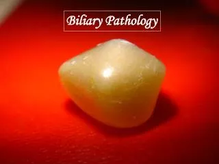
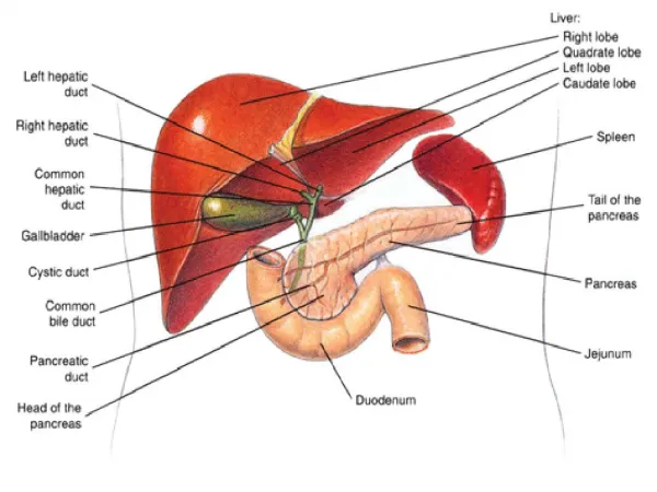
Gallbladder I. cholelithiasis (gallstones) - prevalence … 10% - gallstones: cholesterol x pigment x mixed - 1. cholesterol stones (crystalline cholesterol monohydrate): - supersaturated bile with chol. + nucleation (mikroprecipitates of Ca salts) + stasis - age, women, pregnancy, obesity, hyperlipidemia - pale yellow, large, solitary x multiple (faseted surface) - 2. pigment stones (bilirubin Ca salts): - chronic hemolytic syndromes + biliary infection - black, small, multiple - asymptomatic (70-80% pts.) x severe „colicky“ pain complications: - cholecystitis - hydrops - biliary enteric fistula (duodenum) – gallstone ileus x perforation – diffuse peritonitis - jaundice (biliary tree obstruction) - acute pancreatitis
Cholecystitis pain, fever, nausea 1. acute calculous cholecystitis (stones present) - enlarged GB, fibrin on serosa, stone in neck - pus in lumen (empyema of GB), gangrenous cholecystitis - Mi: inflammation in the wall 2. acute acalculous cholecystitis (stones absent) - postoperative state, trauma, burns, sepsis 3. chronic cholecystitis (stones present) - from acute cholecystitis x de novo - recurrent attacks - mucosal ulcerations + wall fibrosis / inflammation
Extrahepatic bile ducts I. biliary atresia(see above) II. choledocholithiasis (stones in biliary tree) - from GB x primary III. cholangitis - complication of choledocholithiasis, ascending (Gram- rods) - suppurative - liver abscesses Liver abscess bacteria, fungi, parasites (Echinococcus) source: portal vein x hepatic artery x biliary tree complication: perforation – diffuse peritonitis
Cholesterolosis • common • “strawberry gallbladder“
Neoplasms I. carcinoma of GB - women, 7th decade, cholelithiasis (+ chronic cholecystitis) - clinically silent – advanced stage - G: exophytic x infiltrating, neck of GB - Mi: adenocarcinoma / squamous differentiation - liver / bile duct invasion + LN + peritoneum, GIT, lung - 5-year survival: 1% II. carcinoma of EH bile ducts (less common) - older men, PSC - jaundice - G: firm gray nodule in wall x infiltrative - Mi: adenocarcinoma - Klatskin tumor – right and left hepatic duct bifurcation - ampulla of Vater – „ampulloma“
Pancreas I. cysts / cystosis II. cystic fibrosis III. regressive changes (atrophy) IV. pancreatitis V. neoplasms
Cystic fibrosis • most common AR disorder in whites • carrier frequency 1 in 30 • prevalence 1 in 2,000 • defect of secretory process of all exocrine glands
Cystic fibrosis • defective CFTRs defect of chloride ions transport across epithelium epithelium impermeable to chloride ions dehydrated viscid mucus with increased content of NaCl • CFTR gene (7q31-32), 300 mutations • 70% patients: δF508
Cystic fibrosis • GIT - pancreas (80% patients) • viscid mucus in dilated ducts • atrophy (Langerhans islets spared) • fibrocystic disease • malabsorption of fat + vitamins A, D, E, K
Cystic fibrosis • GIT - small GIT glands • newborn: viscid mekonium obstruction of small bowel rupture peritonitis • GIT - bile ducts • secondary biliary cirrhosis • male reproductive tract – vas deferens • infertility (95% males)
Pancreatitis 1. acute interstitial non-purulent pancreatitis - viral inflammation - mumps 2. acute interstitial purulent pancreatitis - rare - ascending x hematogenous 3. acute hemorrhagic pancreatitis / necrosis