Download

1 / 51
510 likes | 610 Vues
Detailed insights on psoriasis and lichen planus, including causes, clinical pictures, histopathology, morphological patterns, clinical varieties, treatment options, and pathogenesis.

E N D
Papulo-squamous diseases Dr.MOHAMED NASRLecturer Of Dermatology & VenereologyZagazig University
PSORIASIS • It is a chronic proliferative skin disease that affects about 1-2% of general population.
Cause: Genetic predisposition with triggering factors: • Trauma: can elecit (koebner phenomenon). • Weather: worse in winter. • Endocrinal factors: It may improves with pregnancy and exacerbate with puberty and menopause. • Infection: steptococcal infection may be followed by guttate psoriasis . • Drugs: ß adrenergic blockers, NSAIDs and ACE inhibitors. • Emotional stress. • Alcoholandsmoking.
Histopathology Epidermis: • Parakeratosis (accelerated incomplete keratinization of horny cells with retension of neuclei) with focal orthokeratosis. • Absent granular cell layer. • Regular acanthosis and elongation of club shaped rete ridges. • Suprapapillary thinning of epidermis. • Munro microabscesses formed of collected neutrophils in stratum corneum or just beneath it. • Spongiform pustules in malpighian layer.
Dermis: • Elongation and edema of dermal papillae. • Dilated tortuous capillaries in the upper portion of papillae. • Perivascular mononuclear and neutrophil inflammatory infiltrate in upper dermis.
Clinical picture: • The primary lesion is a salmon pink papulecovered with silvery scales. Papules may enlarge or coalesce forming plaques. • Sites: extensor surfaces of limbs, elbows, knees, scalp, and nails.
Grattage test • Scraping of psoriatic lesion with the edge of a glass slide results in removal of the scales layer after layer with accentuation of the silvery appearance until a smooth glossy red membrane is finally left. On scratching this membrane pinpoint hemorrhages appear (Auspitz's sign).
Morphological patterns 1-Punctate psoriasis (pin point size) 2-Guttate psoriasis (size of drops) 3-Discoid psoriasis (coin shaped) 4-Annular psoriasis (ring shaped lesions produced by involution of center of the lesions) 5-Geographical psoriasis (curved patterns produced on a large area as the back). 6-Linear psoriasis
Clinical varieties - Flexuralpsoriasis: as in axillae and groins. - Psoriasisof palms and soles: may be either; Typical scaly plaques Or thick, fissured plaques similar to hyperkeratotic eczema Or pustular type - Erythrodermictype: generalised erythema and scaling. - Psoriasisof nails: pitting, yellowish discolouration, circular area of discolouration (oil drop) transverse ridging, onycholysis (distal separation), thickening, subungual hyperkeratosis. - Arthropathic psoriasis. - Psoriasisof scalp: not crossing the hair line (D D from seborrheic dermatitis). - Pustularpsoriasis: localised or generalized, the primary lesions here is a sterile pustule.
Pathogenesis: • Epidermal keratinocyte hyperproliferation. The transit time of epidermal cell maturation from a basal cell to be a horny cell in normal skin is about 26-28 days whereas in psoriatic skin it is only 3-4 days. • T-cell mediated inflammatory processinvolving mainly Th1 cells.
Koebner phenomenon: • It is development of isomorphic pathologic lesions in the traumatized uninvolved skin of patients who have psoriasis. • Psoriasis, warts, lichen planus, vitiligo, molluscum contagiosum and pityriasis rubra pilaris (PRP).
Treatment: A-Topical treatment 1-Tar :crude coal tar 2-5%. 2-Anthralin 0.1-1%. 3-Salicylic acid (3-5%) keratolytic remove scales. 4-Corticosteroids ointment. 5-Intralesional corticosteroid injection of triamcinolone acetonide in localised resistent plaques but less effective in nail psoriaisis. 6-Calcipotriol ointment (vitamin D analogue). It inhibits keratinocyte proliferation.
B-Systemic treatment 1-Methotrexate 2.5mg tab (5mg every 12 hour for 3 doses) every week. It is indicated in erythrodermic psoriasis and generalized pustular psoriasis. Liver function should be monitored. 2-Retinoids;etretinate: 1 mg/kg/day and acitretin 0.5-1 mg/kg/day in severe cases and erythrodermic psoriasis 3-Photochemotherapy(PUVA): psoralens 0.6 mg/kg 2 hours before the exposure to UVA rays 2-3 times weekly 4-Phototherapy: narrowband UVB rays (NBUVB) 5-CyclosporinA: immunosuppresive for severe cases 2.5-5mg/kg/day.
LICHEN PLANUS • Lichen planus is an itchy chronic inflammatory disease which affects skin and mucous membranes.
Pathogenesis: 1-Genetic predisposition 2-Autoimmune 3- Strong association with hepatitis C infection 4- Emotional stress
Clinical picture • The primary lesion is a pruritic shiny violaceous flat topped polygonal papule which retains the skin lines and shows white streaks (wickham’s striae). These striae can be seen well with a magnifying lens. • It is commonly seen on wrist, back of hands, shin and ankles in hypertrophic type, lumbar region, glans penis in annular type and palms and soles. • Mucous membrane lesions are very common (in 30-70% of cases) and may occur alone without skin involvement. • After disappearance of the lesions deep pigmentation is left for several months
Clinical varieties: 1- Atrophic lichen planus; fading annualr or hypertrophic type. 2- Hypertrophic Lichen planus: deep violet rough papules over shin of tibia and ankles. 3- Linear lichen planus. 4- Annular lichen planus (on glans penis). 5- Lichen planus bullosus. 6- Lichen planus circinatus. 7- Lichen plano-pilaris: follicular papules which result in cicatricial alopecia. 8- Lichen planus actinicus: on sun exposed areas in the form of annular lesions with violet brown centre and well defined hypopigmented margin. 9- Lichen planus of mucous membrane: (White streaks or network on the buccal mucosa, fixed white plaques on the tongue or ulcerative lesions). 10- Lichen planus of palms and soles: firm rough non itchy deep yellowish papules. 11- Lichen planus of the nails: thinning of nail plate, linear ridges and grooves, adhesions between dorsal nail fold and nail bed may cause partial destruction of nail (pterygium).
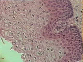
Histopathology: 1-Compact hyperkeratosis. 2-Focal hypergranulosis. 3-Irregular acanthosis and irregular elongation of rete ridges giving a (saw toothed appearance). 4-Liquefactive degeneration of basal layer. 5-Band like inflammatory infiltrate.
Treatment: 1-Assurance and avoidance of stress 2-Topical therapy: a- Fluorinated topical steroid ointment b- Topical tacrolimus c- Intralesionalsteroids (Hypertrophic LP and lichen planus of nail)
3-Systemic therapy: a-Antihistamines b-Systemic steroids (15-20mg/day for 6 weeks) indicated in: 1-severs pruritic or generalized cases 2-ulcerative mucous membrane lesions 3-progressive nail destruction 4-extensive lichenplanopilaris to prevent cicatricial alopecia c- Retinoids d- Cyclosporin A e- Antimalarials for actinic L.P f- NBUVB and PUVA
PITYRIASIS ROSEA • It is an acute self limited disorder characterized by superficial, scaly lesions on the trunk. • It is more common in spring and autumn.
Clinical picture: 1- The primary lesion is the herald patch. 2- It is followed after 5-15 days by multiple similar macules or patches. 3- The long axes of lesions follow lines of cleavage parallel to the ribs in a Christmas tree pattern on the upper chest and back. 4- The eruption fades within 4-8 weeks.
Types: 1- Classical type. 2- Inverted type. 3- Abortive type. 4- Localised type.
Etiology: a- Infective agent: virus (HHV6-7). b-Drug induced (pityriasiform drug eruption) as captopril, metronidazole, ketotifen and barbiturates. c- Autoimmune disease.
Treatment: 1-Reassurance 2-Antihistaminics 3-Calamine lotion 4-Narrow band UVB
PITYRIASIS RUBRA PILARIS • This is a chronic disease characterized by follicular hyperkeratosis, branny scales, orange red erythema and palmoplanter keratoderma.
Etiology: Unknown. • It may be dominantly inherited. • Vitamin A deficiency is also reported.
Clinical signs: 1-Diffuse scaliness of scalp. 2- Patches showing keratotic follicular papules. 3- Dry scaly erythematous areas simulating psoriasis. 4- Hyperkeratosis of palms and soles. 5- Follicular hyperkeratosis of proximal phalanges of fingers and toes. 6- Nails: dull, thickened rough dystrophic. 7-Erythroderma with islands of normal skin.
Types: • Classical adult type (most common 55%) • Atypical adult type (5%) • Classical jeuvenile type (10%) • Circumscribed jeuvenile type (25%) • Atypical jeuvenile type (5%)
Treatment: 1-Emollients in cases of erythroderma to reduce scaling and restore skin barrier 2-Topical steroids and salicylic acid ointment 3-Topical vitamin D analogues (calcipotriol) 4-Oral Vit A: 150,000-300,000 IU/day 5-Isotretinoin and acitretin 6-Methotrexate 10-25mg/week 7-Narrow band UVB 8-PUVA alone or/and oral retinoid