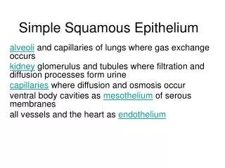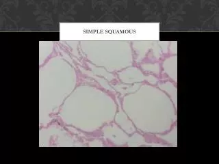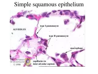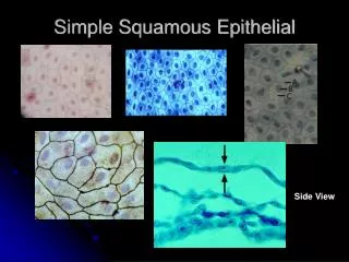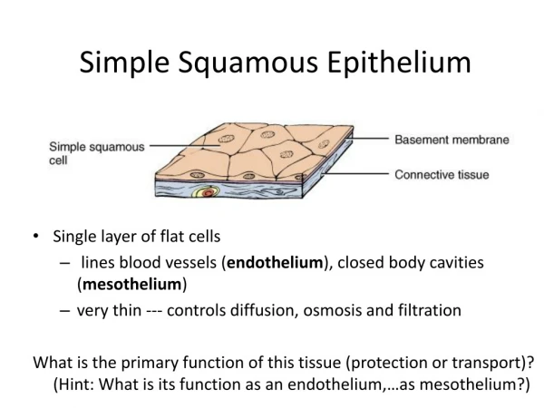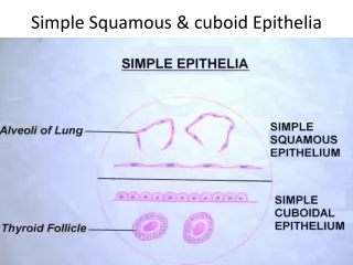
Simple Squamous
Simple Squamous. Simple Cuboidal. Simple Columnar. Stratified Columnar. Pseudostratified Columnar. Loose Connective Tissue. Fibrous Connective Tissue. Cartilage. Bone. Adipose. Skeletal Muscle. Smooth Muscle. Cardiac Muscle. Neuron. Sponges.
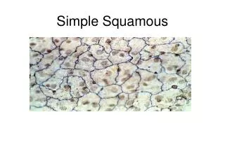

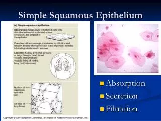
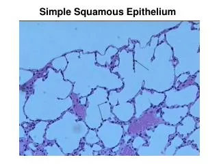
Simple Squamous
E N D
Presentation Transcript
Sponges Sponges are sessile animals that are made up of a loose aggregate of cells which means they are different from other animals because they have no true tissues. They have a cellular-level of organization and the individual cells retain a large degree of independence. The word porifea means “pore-bearers” because a sponge is basically a sac that is full of holes. Sponges are usually classified by their canal systems (with flagellated cells called choanocytes) and the type of skeletal structures they possess.
Body Types and Skeletal Structures Sponges have a large central cavity called a spongocoel. This cavity opens to the outside by a large opening called an osculum. Sponges have three body types depending on the location of their choanocytes: Asconoid: flagellated spongocoels Syconoid: flagellated canals Leuconoid: flagellated chambers The skeletal structures in sponges are called spicules (made of calcium carbonate or silica) and spongin (made up of protein).
Be able to identify the following structures under the microscope: Ostia Incurrent Canal Prosopyle Radial Canal Apopyle Spongocoel Osculum Sponge Anatomy
Cnidaria are separated from other animals because of their radial symmetry. These animals are said to have a tissue-level of organization. They are said to be diploblastic because they have a true outer epidermis and an inner endodermis separated by mesoglea. The body plan for this group is a sac that surrounds a gastrovascular cavity. These organisms are polymorphic and demonstrate two body types in their life cycles (the polyp and the medusa). These organisms all possess nematocysts (stinging cells) that are used to capture prey and for protection).
You need to be able to identify the following structures: tentacles, mouth, gastrovascular cavity, epidermis, gastrodermis, mesoglea and basal disc. Phylum Cnidaria: Hydra
Sexual Reproduction: Ovaries and Testes Asexual Reproduction: Budding Phylum Cnidaria: Hydra Reproduction
Medusa Polyp: Phylum Cnidaria: Obelia
Phylum: Ctenophora The word Ctenophora means “comb-bearer”. They contain comb plates with cilia for movement and tentacles that contain colloblasts to capture their prey.
Platyhelminthes are different from other animals because of there is no space between the gastrovascular cavity and the muscles so they are said to be acoelomates. They are also the first animals that demonstrate bilateral symmetry, which allows these organisms to develop a head with specialized sense organs. These animals are said to have an organ system level of organization. They are said to be triploblastic because they have a true outer epidermis and an inner endodermis separated by a third layer called the mesodermis. The body plan for this group is a solid mass of tissue that surrounds that surrounds a gastrovascular cavity.
Class: Turbellaria These flatworms have eyespots called ocelli that are used for light detection. They have bumps on the side of their head called auricles used as a chemical detectors.
Class: Turbellaria Know the following structures: Pharynx Mouth Gastrovascular Cavity Ocelli Auricles Intestines Anterior Posterior
Class: Trematoda The flukes are flatworms which are parasites that have multiple hosts. Many species spend part of their life cycle in invertebrates and vertebrates such as snails, crabs, fish, birds, etc. They have an outer tegument to protect them from their host.
Class: Trematoda Know the following structures: Oral Sucker Ventral Sucker Esophagus Intestine Testes Ovaries Uterus Shell Gland Yolk Gland
Class: Cestoidea These flatworms are endoparasitic parasites called tapeworms. They have specialized body parts:a head called a scolex and body segments called proglottids.
Know where the following cross sections were taken Anterior: Pharyngeal Posterior Class: Turbellaria Cross Sections
Class: Cestoidea Know the following structures: Scolex Hooks Rostellum Suckers Proglottids Uterus Yolk Gland Testes Ductus deferens Genital Pore Vagina
Phylum: Rotifera The rotifers are animas that exhibit a pseudocoelomate body plan. They are one of the early animals to exhibit an alimentary canal (which has both a mouth and an anus). They exhibit an organ-system level of organization and they aretriploblastic.The word rotifer means wheel bearer because they have jaws and a crown of cilia.
Phylum: Nemertea The ribbon or proboscis worms are animals that are different from other animals because they exhibit an acoelomate body plan but have a fluid sac that some suggest may be an early coelom. They have an alimentary canal, closed circulatory system and the fluid sac mentioned above.
Phylum: Nematoda The nematodes are animals that exhibit a pseudocoelomate body plan. They are one of the first animals to have an alimentary canal (which has both a mouth and an anus). They exibit an organ-system level of ogranization and they are triploblastic. The muscles of nematodes are all longitudinal so they demonstrate a snake-like movement.
Ascaris lumbricoides The human intestinal roundworm may actually be found living as a parasite in the intestines of horses, pigs, and humans. Children that play in the dirt often ingest the eggs. The body is long, slender, smooth, unsegmented and pointed at both ends and lives in the hosts small intestine. The males of this species are about 6 to 10 inches long and have a curved posterior end that bears bristle-like copulatory spicules near the genital pore. The females are about 12 to 14 inches long are not curved near the genital pore.
Necator americanus The American hookworm lives in warm climates because the larvae form is found in the soil and can’t survive colder climates. The adult male is 7- 9mm long and the female adult is 9 – 11 mm long. The adult is found in the small intestines of the host. The eggs are passed in the feces and the juveniles live in the soil until they can burrow into the skin of the host and work their way back into the intestines via the lungs. Heavy infestations can cause anemia or death. Males have conspicuous copulatory bursa supported by fleshy rays.
Trichinella spiralis The pork roundworm is a parasite that infects pigs, rats, humans, and other mammals that are carnivorous. It causes the lethal disease trichinosis. Adult worms penetrate the small intestine where the adult female produces living young. The juveniles burrow into the circulatory system and are carried throughout the body and eventually burrow their way into skeletal muscle and form a cyst. The organism enters the host when a host ingests raw or undercooked meat.
Enterobius vermicularis The pinworm is a common intestinal parasite that infects children of all nations and social classes. The female worm migrates to the anal region and night and deposits her eggs. This causes an irritation around the anus causing it to itch. Scratching the area, may transfer the eggs to the hands which can than be swallowed and a person than is reinfected. Be able to recognize this species (It has a clear tail with the anus at the end of the worm).