Download
1 / 109
1.16k likes | 1.48k Vues
DISEASE OF THE CARDIOVASCULAR SYSTEM. Prof: Tian Dong Ping 田东萍 Department of Pathology , Shantou University Medical College. Section 1 Arteriosclerosis. Introduction:

E N D
DISEASE OF THE CARDIOVASCULAR SYSTEM Prof: Tian Dong Ping 田东萍 Department of Pathology , Shantou University Medical College
Section 1 Arteriosclerosis • Introduction: • Arteriosclerosis encompasses any condition of arterial vessels that result in a thickening or hardening of the walls,It generally includes three condition: • Arteriolosclerosis • Athrosclerosis • Monckeberg`s Sclerosis(medial calcification)
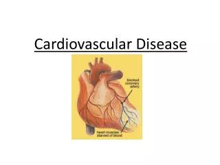
Atherosclerosis Definition: Atherosclerosis is a disease which affects large and medium-size arteries,Characterized by intimal fatty deposits followed by proliferation of smooth muscle cells, ultimately with collagenization. Causes : Not Clear
Clinical Significance • Cause 50% or more deaths. Involved arteries supply brain, heart, kidneys, lower extremities and small bowel. • Myocardial infarct (heart attack). • Cerebral infarction (stroke), and • Aneurysm are major consequences. • Gangrene of lower legs, ischemic heart d/s and ischemic encepholopathy and others
Morphology in Pathology • Three stage and then complicated lesion • Fatty streaks • Location: include the abdominal aorta and large arteries of the lower limb, the coronary arteries e and the circle of Willis. • Grossly :Pale yellow lines running parallel to one another downward for several centimeters, Eath Streak is only 1to 2mm wide.
LM: The lipid is found 1) on endothlial cells overlying the streaks 2) within foam cell The foam cells arise from 1 )lipid-containing smooth muscle cells2) Macrophages
2 Fibrous plaque • The fatty material accumulates to form a fatty pool at the center of the developing plaque. • Overgrowth of fibrous tissue around the fatty pool –fibrous plaque. • Grossly: Pale yellow--- palest, slightly alevated plaque • LM: a great deal foamy cells under the fibrous tissue .
3 Atheromatous plaque ( atheroma) Groosly: different size and number lesion on the surface of the endothelial . It including thick yellow porridge –like material, Sudan III staining the fat shows up. LM:the lumen is lined by fibrous tissue , below which is pink amorphous material.(necrosis)---cholesterol clefts, foamy cell can been see around the edge of lesion. The deposits of calcium which stain blue.
D. Complication of atheromatous plague 1. Calcification 2. Plague ulceration 3. Intimal hemorrhage 4. Thrombosis and embolism 5. Aneurysm
Risk Factors • Age: 4-6th decades; but, the disease begins much younger age, slowly progressive. • Sex: Men are more prone to have the d/s. • After menopause, female increase and by 60-70 yrs equal to male. • Genetic: hyperlipidemia, hypertension, diabetes, smoking and familial risk factor.
Hyperlipidemia • Hyperclosterolemia-more responsible than hypertriglyceridemia for atherosclerosis. • High LDL cause higher risk; HDL acts in reverse, helps prevent atherosclerosis and ischemic heart disease (HDL move choles-terol to liver for excretion in bile). • Genetic defects(familial hyperchlosterole- mia)-inadequate hepatic uptake of LDL.
Pathogenesis • Two major hypotheses: • 1. Response to injury- chronic endothelial injury; insudation of lipoprotein-LDL; adhesion of blood monocytes & platelets; porliferation/migration of smooth muscle cells; lipids accumulation within cells. • 2. Chronic inflammatory response.
Aneurysms and Dissection • Localized abnormal dilatation of a blood vessel, most commonly-aorta & heart. • Blood enters the wall of the artery, dissect- ing between its layers. Marfan • Two major causes for aneurysm-atheroscl- erosis and cystic medial degeneration. • Mycotic aneurysm-infection
Syphilitic (Luetic) Aneurysms • Tertiary stage of syphilis-cardiovascular and nervous systems; obliterative endar-teritis-involved small vessels(vasa vasorum) of the aorta-weaken the wall-aneurysmal formation and scarring of the intima-tree-barking. • Luetic aortitis-aortic valve ring dilatation.
Clinical Course • Rupture-abdominal cavity/retroperitoneal. • Occlusion of a branch vessel at the origin. • Embolism from the atheroma or thrombus. • Impingement on adjacent structure. • Presentation as an abdominal mass. * Rupture is the most feared consequence. The bigger the higher the risk of rupture.
Section 2 • Coronary Atherosclerosis • And Coronary Heart Diseas(CHD)
Coronary Atherosclerosis (CA) • Location: • left coronary artery----the most frequent site of CA • And then the right coronary artery
2.Coronary Heart Diseas(CHD) • Definition: • Cardiac disease resulting from coronary Atherosclerosis and its complications is referred to as Atherosclerotic coronary heart disease • More than 90% is due to atherosclerotic coronary arterial obstruction (Coronary heart disease
Types: *angina pectoris *myocardial infarction *myocardial fibrosis (chronic ischemic heart disease) *Sudden coronary death
angina pectoris • Clinical feature: is paroxysmal pain in the chest, occasionally radiating down the medial aspect of the left arm. • Cause : coronary atherosclerosis, with narrowing or occlusion of the coronary arteries---- oxygen deficiency of the myocardium. • Classically, angina is precipitated by activities increasing myocardial oxygen demand, such as exercise, and is relieved with in minutes by rest or nitroglycerin
2. Myocardial Infarction Cause: Coronary atherosclerosis→ Prolonged ischemia of over 30 to 45 minutes→ muscle death. Location:almost occur in the left ventricle. The most frequent site is the anterior region of the left ventricle, including the anterior two thirds of the interventricular septum.
Grossly: (1) during the first few hours: is not striking (2) 48 to 72 h: paler and drier than normal——“ Coagulation ncrosis”. (3) about 8 to 10 days: a reddish purple zone is noticed at the periphery of the infarct as a result of the granulation tissue, and the infracted area shrinks somewhat because of the resortion of necrotic muscle. (4) about 2 to 3 months: a gray- white or white fibrous shrunken scar replaces he resorbed dead muscle.
LM: (1) 6-12 h- fibres show degenerative changes ① Increased eosinophilia ② Swelling and inflammatory response (2) 48 to 72h: Tissue degradation and removal of necrotic fibers begins. (3) about the third week: scar formation begins (4) By the sixth week: the scar well established
Outcomes: Myocardial infarction depresses ventricular function as a result of the loss of contractility in the necrotic muscle.
Complications and causes of death (1) papillary muscle dysfunction (2) Mural thrombosis and embolism. (3) Rupture of heart. (4) Cardiac Aneurysm. (5)Immediate mortality
3. Myocardial fibrosis Focal fibrosis of the myocardium is the lesion with a chronic, progressive type of myocardial ischemia. The myocardial lesion is observed in the heart of patients who have had a history of attacks of angina pectoris.
Section 3 Hypertension • Most important risk factor in coronary heart disease and cerebrovascular accidents. • Also lead to cardiac hypertrophy-heart failure; aortic dissection; renal failure. • 90-95% are idiopathic and apparently primary (essential), • 5-10% secondary to renal disease and others. • 5% of patients-malignant hypertensive and die within 2 yrs if untreated (diastolic-120mm Hg+)
Definition: Hypertension is the persistent elevation of systemic arterial pressure above normal level. 1. Normal blood pressure: B. P.≤18.6/12 kPa (140/90mmHg) 2. Hypertension: B. P. ≥ 21.3/12.6 kPa (160/95mmHg) 3. Borderline: between normal and hypertension.