Download
1 / 26
320 likes | 715 Vues
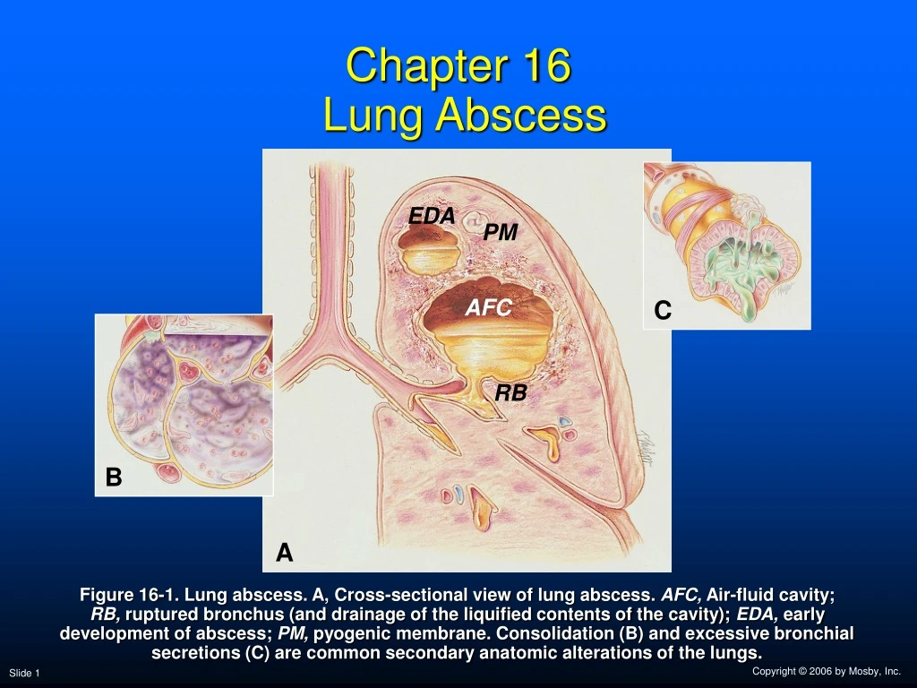
Chapter 16 Lung Abscess. EDA. PM. AFC. C. RB. B. A.

E N D
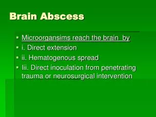
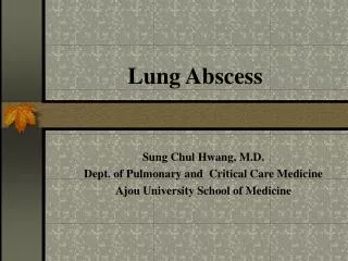
Chapter 16 Lung Abscess EDA PM AFC C RB B A Figure 16-1. Lung abscess. A, Cross-sectional view of lung abscess. AFC, Air-fluid cavity;RB, ruptured bronchus (and drainage of the liquified contents of the cavity); EDA, early development of abscess; PM, pyogenic membrane. Consolidation (B) and excessive bronchial secretions (C) are common secondary anatomic alterations of the lungs.
Anatomic Alterations of the Lungs • Alveolar consolidation • Alveolar-capillary and bronchial wall destruction • Tissue necrosis • Cavity formation • Fibrosis and calcification of the lung parenchyma • Bronchopleural fistulae • Atelectasis • Excessive airway secretions and empyema
Etiology • Pneumonia caused by aspiration (most common) • Klebsiella • Staphylococcus • Predisposing factors for aspiration • Alcohol abuse • Seizure disorders • General anesthesia • Head trauma • Cerebrovascular accident • Swallowing disorders
Etiology (Less frequent causes) • Aerobic organisms • Streptococcus pyogenes • Klebsiella pneumoniae • Escherichia coli • On rare occasions • Streptococcus pneumoniae • Pseudomonas aeruginosa • Legionella pneumophila
Etiology (Other organisms that may lead to a lung abscess) • Mycobacterium tuberculosis • Fungal organisms • Histoplasma capsulatum • Coccidioides immitis • Blastomyces • Aspergillus fumigatus • Parasites • Paragonimus westermani • Echinococcus • Entamoeba histolytica
Etiology Lung abscess may also develop from: • Bronchial obstruction • Vascular obstruction • Interstitial lung disease • Bullae or cysts • Penetrating chest wounds
Overview of the Cardiopulmonary Clinical Manifestations Associated with LUNG ABSCESS The following clinical manifestations result from the pathophysiologic mechanisms caused (or activated) by Alveolar Consolidation (see Figure 9-8), and when the abscess is draining, by Excessive Bronchial Secretions (see Figure 9-8)—the major anatomic alterations of the lungs associated with chronic bronchitis (see Figure 16-1).
Clinical Data Obtained at the Patient’s Bedside Vital signs • Increased respiratory rate • Increased heart rate, cardiac output, blood pressure
Clinical Data Obtained at the Patient’s Bedside • Chest pain/decreased chest expansion • Cyanosis • Cough, sputum production, and hemoptysis • Chest assessment findings • Increased tactile and vocal fremitus • Dull percussion note • Bronchial breath sounds • Diminished breath sounds • Whispered pectoriloquy • Pleural friction rub
Figure 2-11. A short, dull, or flat percussion note is typically produced over areas of alveolar consolidation.
Figure 2-16. Auscultation of bronchial breath sounds over a consolidated lung unit.
Figure 2-19. Whispered voice sounds auscultated over a normal lungare usually faint and unintelligible.
Clinical Data Obtained from Laboratory Tests and Special Procedures
Pulmonary Function Study: Expiratory Maneuver Findings FVC FEVT FEF25%-75% FEF200-1200 N or N or N PEFRMVV FEF50% FEV1% N N or N N or
Pulmonary Function Study: Lung Volume and Capacity Findings VT RV FRC TLC N or VC IC ERV RV/TLC% N
Arterial Blood Gases Mild to Moderate Lung Abscess • Acute alveolar hyperventilation with hypoxemia pH PaCO2 HCO3- PaO2 (Slightly)
Time and Progression of Disease Disease Onset Alveolar Hyperventilation 100 90 Point at which PaO2 declines enough to stimulate peripheral oxygen receptors 80 70 60 PaO2 PaO2 or PaCO2 50 40 30 PaCO2 20 10 0 Figure 4-2. PaO2 and PaC02 trends during acute alveolar hyperventilation.
Arterial Blood Gases Severe Lung Abscess • Acute ventilatory failure with hypoxemia pH PaCO2 HCO3- PaO2 (Slightly)
Time and Progression of Disease Disease Onset Alveolar Hyperventilation Acute Ventilatory Failure 100 Point at which disease becomes severe and patient begins to become fatigued 90 Point at which PaO2 declines enough to stimulate peripheral oxygen receptors 80 70 PaCO2 Pa02 or PaC02 60 50 40 30 PaO2 20 10 0 Figure 4-7. PaO2 and PaCO2 trends during acute ventilatory failure.
Oxygenation Indices QS/QT DO2 VO2 C(a-v)O2 Normal Normal O2ER SvO2
Abnormal Laboratory Testsand Procedures Sputum examination • Gram-positive organism • Streptococcus • Anaerobic organisms • Peptococcus • Peptostreptococcus • Bacteroides • Fusobacterium
Radiologic Findings Chest radiograph • Increased density • Cavity formation • Cavity with air-fluid levels • Fibrosis • Pleural effusion
Figure 16-2. Reactivation tuberculosis with a large cavitary lesion containing an air-fluid level in the right lower lobe. Smaller cavitary lesions are seen in other lobes. (From Armstrong P et al: Imaging of diseases of the chest, ed 2, St. Louis, 1995, Mosby.)
General Management of Lung Abscess Respiratory care treatment protocols • Oxygen therapy protocol • Bronchopulmonary hygiene therapy protocol • Hyperinflation therapy protocol
General Management of Lung Abscess Medications and procedures commonly prescribed by the physician • Antibiotics • Surgery