Download
1 / 46
460 likes | 712 Vues
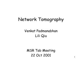
Tomography of flagellar pocket. Lacomble et al., J Cell Sci. 122:1081-90. Sleeping Sickness and Trypanosomes. Trypanosomias. Trypanosomes were first described in frogs 1855 Griffith Evans identifies T. evansi as agent of surra (a horse and camel disease) in 1880

E N D
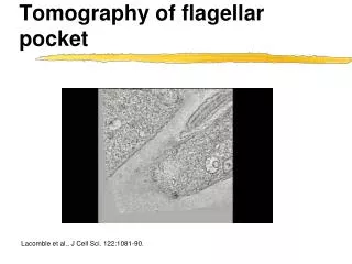
Tomography of flagellar pocket Lacomble et al., J Cell Sci. 122:1081-90.
Trypanosomias • Trypanosomes were first described in frogs 1855 • Griffith Evans identifies T. evansi as agent of surra (a horse and camel disease) in 1880 • David Bruce identifies T. brucei as cause of Nagana and demonstrates transmission by Tse-tse flies David Bruce, 1855-1931
The kinetoplast consists of a complex network of concatenated DNA cirlces Mensa-Wilmot lecture will tell you what the circles are for
Tse tse flies • Parasites are taken up with the blood meal (stumpy forms are cell cycle arrested and ‘ready to go’ for the next host • Transformation into procyclic trypomastigotes in the midgut • Migration into the ectoperitrophic space where parasites replicate • Passage into salivary glands, differentiation into epimastiogotes which attach to the epithelium and massively replicate • Transformation into infectious metacyclic trypomastigotes • Again these are cell cycle arrested ‘sleepers’
Trypanosmes have sex and likely it happens in the salivary gland • Genetic exchange occurs (e.g. double drug resistance occurs after coinfection of fly with single resistant parents) • Genetic exchange/sex is likely not obligatory to complete fly development and population genetics suggest “modest” sex • Nobody has seen it, yet it likely involves fusion and meiosis (progeny appears largely diploid but there are also polyploids) • Most likely this exchange/fusion occurs among detached salivary gland epimastigotes
Trypanosmes have sex and likely it happens in the salivary gland GFP & RFP only parents early on in salivary gland Red, green & yellow progeny Red, green & yellow progeny Gibson et al., Parasites & Vectors 2008, 1:4
Insect stages and blood stream forms are very different • Different stages express different sets of surface proteins • Insect forms have large mitochondria with many cristae • Insect stages have an aerobic metabolism and a full respiratory chain • Blood stream forms only engage in glycolysis and excrete pyruvate and glycerol • Note that transmission stages do not replicate and are arrested in development (ladies in waiting)
Several species of trypanosomes cause disease in domestic animals and man • T. brucei rhodesiense & gambiense cause sleeping sickness • T. brucei brucei, T. congolense and T. vivax cause Nagana in cattle • T. equiperdum causes sexually transmitted disease in horses and camels (interestingly, T. equiperdum is a recent ‘petite’ mutant of T. brucei (loss of mitochondrial genome or kDNA) Lai et al. 2008, http://www.pnas.org/content/105/6/1999.full • Loss of oxidative phosphorylation locks parasite into BS form – or the other way around, leaving Africa and tse-tse transmission makes the mitochondrion dispensable • (There are trypanosomes infecting many species of animals and even plants and every single deer in the State of Georgia)
Sleeping sickness in man • Trypanosomes multiply in the tissue around the initial bite site • This often results in a characteristic local inflamation the trypansomal chancre • From there they enter the blood and lymphatic system
Sleeping sickness in man • Enlargement of the lymphatic glands (especially in the posterior triangle of the neck) can be an early sign of the diseasese (Winterbottom sign, not as common in rhodesiense infection). • Aspiration of swollen gland often reveals parasites.
Sleeping sickness in man • Once parasites enter blood stream fever sets in (low and irregular in gambiense and high and periodic in rhodesiense • General toxic symptoms include headache, facial oedema, nausea and vomiting,back and bone pain • Symptoms at this stage are rather mild in gambiense but can be servere in rhodesiense with often fatal outcome
Sleeping sickness in man • The second stadium of trypansomiasis is characterized by progressive anemia and kachexia. • Both features are primarily due to extremely high serum levels of TNFa • TNFawas isolated both as factor with tumor necrotic effect as well as kachexin inducing wasting in nagana
Sleeping sickness in man • In later stages of infection parasites pass the blood brain barrier and infect the CNS • Presence of parasites leads to meningo-encephalitis with progressive neurological involvement, which ultimately ends in coma (sleeping sickness) • Untreated trypanosomiasis is always fatal
Sleeping sickness in man • The progressive encephalitis can cause severe dementia with sometimes aggressive behavior • Disease progression especially CNS invasion is much faster in rhodesiense • Gambiense can take a year or two rhodesiense usually passes the blood brain barrier within a month
Nagana is the major impediment to cattle production in Africa • Almost the entire area of subsaharan Africa which is suitable for cattle is Tsetse infested • High losses due to anemia and cachexia especially in productive breeds
Wild animals are important reservoirs for human and cattle trypanosomiasis
Why is trypanosomiasis so deadly? • Trypanosomes are highly susceptible to antibodies and complement • They live fully exposed to antibodies in the blood stream • They induce a very strong antibody response • Still they manage to thrive in the same host for a year or longer
Why is trypanosomiasis so deadly? • Infection is characterized by periodic waves of parasitemia
Why is trypanosomiasis so deadly? • Infection is characterized by periodic waves of parasitemia • Each wave represents a single antigenically distinct clone or serotype
Antigenic variation • The entire trypanosome population seems antigenically uniform but at a very low frequency divergent (so called switched) serotypes are encountered
Antigenic variation • Trypanosomes are covered with a dense surface coat • Variant specific antisera strongly react with this surface coat • Surface coats from different clones are antigenically distinct
The surface coat consists of a single 65 kDa glycoprotein • A single protein can be labeled on the surface of trypanosomes • Upon parasite lysis this protein becomes soluble and can be purified to homogeneity fairly easily. George Cross http://tryps.rockefeller.edu/
Different antigenic variants have different surface glycoproteins • VSGs from different clonal isolates have the same molecular weight but vastly different amino acid compositions • Vaccination with a given VSG protects against challenge with the homologous isolate but not against another variant.
VSGs share a common structure • All VSGs are 65 kDA glycoproteins • Most contain classical N-linked glycans and all are anchored via a GPI glycolipid (cross reacting determinant) • Two domains can be cleaved by trypsin • The outer domain is highly variable and the only conservation detected is the position of cysteines • VSG forms dimers
Antigenic variation • VSG dimers form a densly packed surface coat • Other (non-variant) proteins like transferrin receptor or hexose transporter are hidden in this coat
Trypansomes harbor ~1000 different VSG genes • The genomic organization of trypanosomes is quite complex with 20 chromosomes and 100 mini chromosomes • Great variability of chromosome size between isolates • 6-10% of the total DNA is coding for VSGs (~1000 genes) • Only one is expressed • 3 very peculiar detailsemerged from studying the mRNA of VSG: all trypanosome mRNAs seem to have the same 5’end, and the VSG mRNA encodes a hydrophobic c-terminus absent from the mature protein sequence, VSG message is transcribed by Pol I
Antigenic variation • mRNA derived from only a single VSG gene can be detected at one time • VSG expression is controlled at the level of transcription initiation • Regulation of promoter activity is used to control gene expression in many organisms
Transcription in trypanosomes is polycistronic • But, only very few promoters have been identified in trypanosomes and they did not seem to regulate the expression of VSG • Also surprisingly transcription in trypanosomes was found to be polycistronic • Polycistronic means that a number of genes are transcribed at the same time into one long messenger RNA • In bacteria this message is translated into protein, in trypanosomes further processing is needed
Transcription is polycistronic • The 39 first (5’) base pairs of all trypanosme mRNAs are identical, this sequence is not found in the genomic locus of these genes • Individual mature mRNAs are derived from large polycistronic transcripts and short SL-RNAs by trans-splicing (details in Mensa-Wilmot lecture) • This might help control – but was shown not to be the key to antigenic variation
VSGs are expressed from telomeric polycistronic expression sites • Active VSG genes are typically at the “ends” of chromosomes (telomeres) • They are found in “expression sites” • Genes are read in (~20) expression sites like tapes in a tape recorder but only one recorder is playing at a time • How do you get a new tape in and how are the recorders controlled e.g. switched on and off?
Antigenic variation • Transposition of VSG genes occurs by intra- or intermolecular recombination • This explains switching but not really why one gene is active and all the others are silent
Antigenic variation • Regulation could be achieved by modification of chromatin • Indeed active and inactive sites differ in the amount of a special modified base called J (b-glucosyl-hydroxy-methyluracil - a T variant) and there are newly discovered differences in histone methylation and acetylation patterns (Bob Sabatini will go over this in detail on Monday)
For the next experiment we need a mushroom Amantia bisporingea, the Destroying Angel http://www.mushroomexpert.com
VSG is transcribed by Pol I • a-amanitin is a specific and highly potent RNA polymerase inhibitor • Cells have specialized RNA polymerases to transcribe different genes • In most cells mRNA which encodes proteins is transcribed by the RNA polymerase Pol2 (this enzyme can be inhibited by the toxin a amanitin) • Ribosomal RNA is generally transcribed by Pol1 (which is resistant to the toxin) • VSG transcription is insensitive to a-amanitin suggesting it is transcribed by the highly processive Pol I (however all other mRNAs for proteins seem to be made using Pol II as everywhere else) • How could this help to explain allelic exclusion? tubulin rRNA VSG Drug
African trypansome cellular architecture Nucleus Nucleoulus Kinetoplast
How is a single expression site activated? • Location, location, location • PolI antibody detects two spots in blood stream forms: the nucleolus (where rRNA is made) and a second locus outside of the nucleolus Navarro M, Gull K. Nature 414:759-63
How is a single expression site activated? • The additional spot of PolI is not the nucleolus Navarro M, Gull K. Nature 414:759-63
How is a single expression site activated? • The extranuclear PolI structure is transcriptionally active control a-amanitin a-amanitin Navarro M, Gull K. Nature 414:759-63
How is a single expression site activated? • Labeling of the expression sites using GFP-Lac • Active, not inactive VSG expression sites colocalize with the extranuclelarPolI spot active VSG inactive VSG Navarro M, Gull K. Nature 414:759-63
Antigenic variation • Only a single VSG gene out of ~1000 is expressed • Expression occurs out of teleomeric expression sites (the tape recorder) • To switch genes on they are transposed into an active expression site by several mechanisms • Expression seems promoter independent • Inactive DNA is modified • Expression seems to be controlled by physical association of ES with a single POL1 transcription particle per nucleus