Diencephalon, Brain Stem and Cranial Nerves
160 likes | 447 Vues
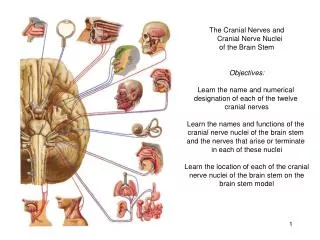
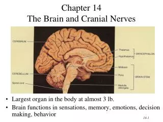
Diencephalon, Brain Stem and Cranial Nerves. Day 5 Pages: 242-246. Diencephalon. Located between the cerebral hemispheres and above midbrain Composed largely of gray matter Surrounds 3 rd ventricle Includes: Thalamus Hypothalamus Optic tracts Optic chiasma Infundibulum

Diencephalon, Brain Stem and Cranial Nerves
E N D
Presentation Transcript
Diencephalon, Brain Stem and Cranial Nerves Day 5 Pages: 242-246
Diencephalon • Located between the cerebral hemispheres and above midbrain • Composed largely of gray matter • Surrounds 3rd ventricle • Includes: • Thalamus • Hypothalamus • Optic tracts • Optic chiasma • Infundibulum • Posterior pituitary gland • Mammillary bodies • Pineal gland
Parts to Diencephalon • Optic tracts/optic chiasma • Formed by optic nerve fibers crossing over each other • Infundibulum • Behind optic chiasma, attachment for pituitary gland • Posterior pituitary gland • Hangs from floor of hypothalamus • Mammillary Bodies • Two rounded structures behind infundibulum • Pineal gland • Cone shaped structure attached to upper portion of diencephalon
Thalamus • Bulge into 3rd ventricle • Central relay station for all sensory impulses except smell (ascending fibers) • Channels impulses to appropriate regions of cortex for interpretation • Can communicate with cerebral cortex by means of descending fibers
Hypothalamus • Located below thalamus and forms floor of 3rd ventricle • Maintains homeostasis and links endocrine to nervous system • Regulates: • Heart rate and arterial BP • Body Temp. • H2O and electrolyte balance • Control of hunger and body weight • Control movements and glandular secretions of stomach and intestines • Production of neurosecretory substances and stimulation of pituitary gland to secrete hormones • Sleep and wakefulness
Other functions of Diencephalon • Limbic System • Comprised of parts of: cerebral cortex, frontal and temporal lobes, hypothalamus, thalamus basal ganglia and other deep masses called nuclei • Controls emotional experiences and expressions • Can modify the way a person acts • Fear, anger, pleasure, and sorrow • Guides persons behavior towards a likely increase in survival.
Brain Stem • Bundle of nervous tissue that connects cerebrum to spinal cord. • Consists of three parts • Midbrain • Pons • Medulla Oblongata
Midbrain • Located at the top between diencephalon and pons • Contain corticospinal tracts which are the main motor pathways between cerebrum and lower parts of nervous system • Contains several masses of gray matter that serve as reflex centers.
Pons • Rounded bulge on underside of brain stem • Dorsal side relays impulses to and from M.O and cerebrum. • Ventral side transmits impulses to cerebrum and cerebellum. • Also relays sensory impulses from PNS to higher brain centers
Medulla Oblongata • End of brain stem • All ascending and descending nerve fibers pass through MO • Controls vital visceral activites • Cardiac center • Alters heart rate • Vasomotor Center • Constricting and dilating of blood vessels • Respiratory Center • Regulate rate, rhythm, and depth of breathing • Also responsible for coughing, sneezing, swallowing and vomiting.
Reticular Formation • Found throughout the brain stem • Network of nerve fibers • Responsible for taking sensory impulses and activating cerebral cortex into a state of wakefulness • Decreased activity in reticular formation is known as sleep. • Comatose state: • Point at which the reticular formation is injured and cannot be aroused even with strong stimulation.
Cerebellum • Large mass of tissue located below occipital bone. • Divided into two hemispheres • Surrounded by cerebral cortex • Communicates with CNS by three pairs of nerve tracts • Cerebellar peduncles
Cellebellar Peduncles • Inferior • Brings sensory information concerning position of limbs, joints, and other body parts • Middle • Transmits signals from the cerebral cortex to the cerebrellum concerning desired positions of these parts. After interpretation/analysis, sends pulses on to 3rd pair • Superior • Incorporated into motor impulses that get sent down brainstem to move body in desired way.
Cerebellum • Reflex center for integrating sensory information concerning position of body parts and coordination of skeletal muscle movements • Maintains posture • Damage/Injury • Tremors • Inaccurate movements of voluntary muscles • Loss of muscle tone • Reeling walk • Loss of equilibrium
Review • What are the major functions of the thalamus? The hypothalamus? • How may the limbic system influence behavior? • List the structures of the brain stem. • What vital reflex centers are located in the brain stem? • What is the function of the reticular formation? • Where is the cerebellum located? • What are the major functions of the cerebellum?