Download
1 / 32
320 likes | 568 Vues
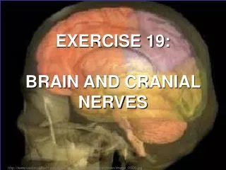
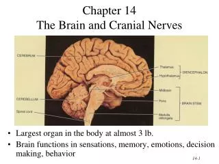
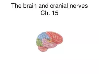
The brain and cranial nerves Ch. 15. Development of the brain. Major parts of the brain. Brain stem Medulla oblongaga, pons, midbrain Cerebellum Diencephalon Thalmus, hypothalamus, and associated structures Cerebrum Largest and most distinctive part of brain.

E N D
Major parts of the brain • Brain stem • Medulla oblongaga, pons, midbrain • Cerebellum • Diencephalon • Thalmus, hypothalamus, and associated structures • Cerebrum • Largest and most distinctive part of brain
Protective structures of brain • Skull • Meninges • Division of brain into hemispheres • Blood-brain barrier • Ventricles
Meninges • Dura mater • Sinuses contain fluid and blood vessels • Arachnoid mater • Covers cranial blood vessels • Pia mater • Attached to surface of brain
Meninges help separate the brain into compartments • Falx cerebri (cerebral hemispheres) • Tentorium cerebelli- separates cerebellum from cerebrum • Falx cerebelli- divides cerebellum into hemipsheres • Diaphragma sellae- lines sella turcica
Distribution of cerebrospinal fluid (CSF) • Ventricles and subarachnoid space • Shock absorber • Chemical balance (electrolytes) • Circulation of nutrients and wastes • Formed in choroid plexuses • Reabsorbed through arachnoid villi
Ventricles • 4 ventricles • CSF circulates between ventricles and spinal canal • 4th ventricle is continuous with spinal cord
Blood supply to brain • No oxygen or energy stores • Internal carotid and vertebral arteries supply oxygenated blood • Drained by internal jugulars
The brain stem: medulla oblongata • White matter: ascending and descending tracts • Pyramids formed by motor tracts; neurons cross from one side to the other • Gray matter (nuclei); cardiovascular, rhythmicity area, various reflexes; cranial nerves
What about the cranial nerves? • 12 pairs • Part of peripheral nervous system (PNS) • Numbered I through XII, in order of anterior to posterior emergence from the brain • Nose- I; eyes- II; inner ear (VIII); brain stem (III-XII); spinal cord (XI) • I and II are sensory; others are mixed • Sensory cell bodies are in ganglia; motor cell bodies are in brain • Motor function is somatic (skeletal) or autonomic
Cranial nerves • I-olfactory • II-optic • III- oculomotor • IV-trochlear • V-trigeminal-opthalmic, maxillary, mandibular • VI-abducens • VII- facial
Eye function is complex • III- eyeball movement as well as intrinsic muscles • IV-superior oblique eye muscle • V-eyelids • VI-lateral movements of eye
Medulla oblongata and associated cranial nerves (inferior view) • Vestibulocochlear (VIII)-hearing • Glossopharyngeal (IX)- taste, chewing, salivation • Vagus (X)-multiple (digestive, heart rate, sensation) • Accessory (XI)-swallowing, head and neck movement • Hypoglossal (XII)-tongue movements during speech
Pons is directly superior to medulla • Nuclei and tracts that connect one part of the brain with another • Pneumotaxic (limits inhalation) and apneustic (prolongs inhalation) centers • Associated with cranial nerves: • Trigeminal (V) chewing • Abducens (VI) movement of eyeballs • Facial (VII)- facial expression; secretion of saliva and tears • Vestibulcochlear (VIII) –balance, equilibrium, hearing
Reflexes and tracts characterize the midbrain (mesencephalon) • Superior and inferior colliculi • Superior- eye reflexes • Inferior- auditory reflexes • substantia nigra • dopaminergic; motor coordination • Red nuclei • Tracts (cerebral peduncles) • Association with cranial nerves: • Oculomotor (III)- movement of eyeball, puil and lens • Trochlear (IV)- movement of eyeball
What does the midbrain look like? Transverse view Posterior view
Reticular formation • Network of cell bodies and myelinated axons • Extends from spinal cord to diencephalon • Helps maintain muscle tone • RAS (reticular activating system) extends to cerebral cortex; helps maintain wakefulness
The cerebellum • Each hemisphere divided into lobes • Anterior, posterior- subconsccious skeletal movement • Flocculonodular- equilibrium • Peduncles organize transmission between cerebellum and rest of brain • Skilled movements • Posture and balance
Functions of cerebellum • Adjusting posture (involuntarily) • Memory of learned movements • Regulates (inhibits) activity in cerebral cortex, basal nuclei, and motor pathways
Diencephalon: thalamus and associated structures • Thalamus is major relay station of sensory information to cerebrum (basal nuclei and cortex) • Anterior (limbic) • Medial • Ventral group • Anterior and lateral (motor) • Posterior (sensory) • Integration of sensory information, perception, emotional state, learning, memory
Hypothalamus • Control of ANS • Hormone production • Regulation of behavior (with limbic system) • Satiety and thirst • Thermoregulation • Epithalamus- pineal gland • Subthalamus-coordination
Cerebrum- most recognizable part of the brain • Left and right hemispheres • Corpus callosum • Lobes formed by sulci and fissures • Insula is deep to the superficial lobes
Basal ganglia • Globus pallidus, putamen, caudate nucleus • (motor) input from cerebral cortex and each other • Control of movement • Damage causes tremor, rigidity, involuntary movements • May intersect limbic system
Limbic system, the emotional brain • Major components: • Amygdala • Hippocampus • Various nuclei and tracts • Olfactory bulbs
Organization of the cerebral cortex • Sensory areas • Generally in parietal or temporal lobe • Primary somatosensory area (postcentral gyrus) • Primary visual area, auditory, gustatory, olfactory • Motor areas • Primary motor area- precentral gyrus • Broca’s area
Association areas • Association areas • Somatosensory-identify sensory input; memory • Visual- recognition, recall • Auditory- what do you hear? • Wernicke’s- what are you saying? • Common integrative area-full integration of sensory input • Premotor- learned motor skills • Frontal eye field area- taking in visual information
Hemispheric lateralization • Each hemisphere controls the opposite side of the body • Corpus callosum connects hemispheres • People tend to concentrate activities in one hemisphere • Left- speech, reasoning, numerical skills • Right- spatial awareness, pattern recognition, musical and artistic sensibility
Summary • Brain grows rapidly during fetal and juvenile development; loses mass with aging • Organization of brain and functional areas is sophisticated with a considerable amount of overlap
Summary • “Higher order” functions in cerebrum • Diencephalon is processing center. Hypothalamus integrates emotions, autonomic, hormone function • Midbrain- visual, auditory, somatic motor • Pons-motor control • Cerebellum-”fine-tunes” • Medulla oblongata- regulates autonomic function