Download

1 / 62
700 likes | 885 Vues
Explore the brain's principal parts, protective coverings, blood supply, cerebrospinal fluid, and key brain regions like the medulla and pons. Learn about cranial nerves and their functions.

E N D
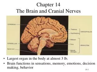
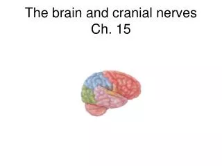
Chapter 14The Brain and Cranial Nerves • Largest organ in the body at almost 3 lb. • Brain functions in sensations, memory, emotions, decision making, behavior
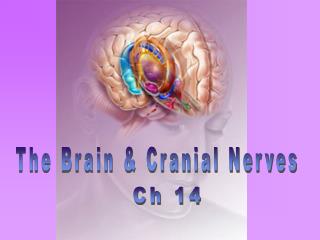
Principal Parts of the Brain • Cerebrum • Diencephalon • thalamus & hypothalamus • Cerebellum • Brainstem • medulla, pons & midbrain
Protective Coverings of the Brain • Bone, meninges & fluid • Meninges same as around the spinal cord • dura mater • arachnoid mater • pia mater • Dura mater extensions • falx cerebri • tentorium cerebelli • falx cerebelli
Blood Supply to Brain • Arterial blood supply is branches from circle of Willis on base of brain (page 699) • Vessels on surface of brain----penetrate tissue • Uses 20% of our bodies oxygen & glucose needs • blood flow to an area increases with activity in that area • deprivation of O2 for 4 min does permanent injury • at that time, lysosome release enzymes • Blood-brain barrier (BBB) • protects cells from some toxins and pathogens • proteins & antibiotics can not pass but alcohol & anesthetics do • tight junctions seal together epithelial cells, continuous basement membrane, astrocyte processes covering capillaries
Cerebrospinal Fluid (CSF) • 80-150 ml (3-5oz) • Clear liquid containing glucose, proteins, & ions • Functions • mechanical protection • floats brain & softens impact with bony walls • chemical protection • optimal ionic concentrations for action potentials • circulation • nutrients and waste products to and from bloodstream
Origin of CSF • Choroid plexus = capillaries covered by ependymal cells • 2 lateral ventricles, one within each cerebral hemisphere • roof of 3rd ventricle • fourth ventricle
Drainage of CSF from Ventricles • One median aperture & two lateral apertures allow CSF to exit from the interior of the brain
Reabsorption of CSF • Reabsorbed through arachnoid villi • grapelike clusters of arachnoid penetrate dural venous sinus • 20 ml/hour reabsorption rate = same as production rate
Hydrocephalus • Blockage of drainage of CSF (tumor, inflammation, developmental malformation, meningitis, hemorrhage or injury • Continued production cause an increase in pressure --- hydrocephalus • In newborn or fetus, the fontanels allow this internal pressure to cause expansion of the skull and damage to the brain tissue • Neurosurgeon implants a drain shunting the CSF to the veins of the neck or the abdomen
Medulla Oblongata • Continuation of spinal cord • Ascending sensory tracts • Descending motor tracts • Nuclei of 5 cranial nerves • Cardiovascular center • force & rate of heart beat • diameter of blood vessels • Respiratory center • medullary rhythmicity area sets basic rhythm of breathing • Information in & out of cerebellum • Reflex centers for coughing, sneezing, swallowing etc
Ventral Surface of Medulla Oblongata • Ventral surface bulge • pyramids • large motor tract • decussation of most fibers • left cortex controls right muscles • Olive = olivary nucleus • neurons send input to cerebellum • proprioceptive signals • gives precision to movements
Dorsal Surface of Medulla Oblongata • Nucleus gracilis & nucleus cuneatus = sensory neurons • relay information to thalamus on opposite side of brain • 5 cranial nerves arise from medulla -- 8 thru 12
XII = Hypoglossal Nerve • Controls muscles of tongue during speech and swallowing • Injury deviates tongue to injured side when protruded • Mixed, primarily motor
XI = Spinal Accessory Nerve • Cranial portion • arises medulla • skeletal mm of throat & soft palate • Spinal portion • arises cervical spinal cord • sternocleidomastoid and trapezius mm.
X = Vagus Nerve • Receives sensations from viscera • Controls cardiac muscle and smooth muscle of the viscera • Controls secretion of digestive fluids
IX = Glossopharyngeal Nerve • Stylopharyngeus m. (lifts throat during swallowing) • Secretions of parotid gland • Somatic sensations & taste on posterior 1/3 of tongue
VIII = Vestibulocochlear Nerve • Cochlear branch begins in medulla • receptors in cochlea • hearing • if damaged deafness or tinnitus (ringing) is produced • Vestibular branch begins in pons • receptors in vestibular apparatus • sense of balance • vertigo (feeling of rotation) • ataxia (lack of coordination)
Injury to the Medulla • Hard blow to the back of the head may be fatal • Cranial nerve malfunctions on same side as injury;loss of sensation or paralysis of throat or tongue; irregularities in breathing and heart rhythm
Pons • One inch long • White fiber tracts ascend and descend • Pneumotaxic & apneustic areas help control breathing • Middle cerebellar peduncles carry sensory info to the cerebellum • Cranial nerves 5 thru 7
VII = Facial Nerve • Motor portion • facial muscles • salivary & nasal and oral mucous glands & tears • Sensory portion • taste buds on anterior 2/3’s of tongue
VI = Abducens Nerve • Lateral rectus eye muscle
V = Trigeminal Nerve • Motor portion • muscles of mastication • Sensory portion • touch, pain, & temperature receptors of the face • ophthalmic branch • maxillary branch • mandibular branch
Midbrain • One inch in length • Extends from pons to diencephalon • Cerebral aqueduct connects 3rd ventricle above to 4th ventricle below
Midbrain in Section • Cerebral peduncles---clusters of motor & sensory fibers • Substantia nigra---helps controls subconscious muscle activity • Red nucleus-- rich blood supply & iron-containing pigment • cortex & cerebellum coordinate muscular movements by sending information here from the cortex and cerebellum
Dorsal Surface of Midbrain • Corpora quadrigemina = superior & inferior colliculi • coordinate eye movements with visual stimuli • coordinate head movements with auditory stimuli
IV = Trochlear Nerve • Superior oblique eye muscle
III = Oculomotor Nerve • Levator palpebrae raises eyelid (ptosis) • 4 extrinsic eye muscles • 2 intrinsic eye muscles • accomodation for near vision (changing shape of lens during reading) • constriction of pupil
Reticular Formation • Scattered nuclei in medulla, pons & midbrain • Reticular activating system • alerts cerebral cortex to sensory signals (sound of alarm, flash light, smoke or intruder) to awaken from sleep • maintains consciousness & helps keep you awake with stimuli from ears, eyes, skin and muscles • Motor function is involvement with maintaining muscle tone
Cerebellum • 2 cerebellar hemispheres and vermis (central area) • Function • correct voluntary muscle contraction and posture based on sensory data from body about actual movements • sense of equilibrium
Cerebellum • Transverse fissure between cerebellum & cerebrum • Cerebellar cortex (folia) & central nuclei are grey matter • Arbor vitae = tree of life = white matter
Cerebellar Peduncles • Superior, middle & inferior peduncles attach to brainstem • inferior carries sensory information from spinal cord • middle carries sensory fibers from cerebral cortex & basal ganglia • superior carries motor fibers that extend to motor control areas
Diencephalon Surrounds 3rd Ventricle • Surrounds 3rd ventricle • Superior part of walls is thalamus • Inferior part of walls & floor is hypothalamus
Thalamus • 1 inch long mass of gray mater in each half of brain (connected across the 3rd ventricle by intermediate mass) • Relay station for sensory information on way to cortex • Crude perception of some sensations
Thalamic Nuclei • Nuclei have different roles • relays auditory and visual impulses, taste and somatic sensations • receives impulses from cerebellum or basal ganglia • anterior nucleus concerned with emotions, memory and acquisition of knowledge (cognition)
Hypothalamus • Dozen or so nuclei in 4 major regions • mammillary bodies are relay station for olfactory reflexes;infundibulum suspends the pituitary gland • Major regulator of homeostasis • receives somatic and visceral input, taste, smell & hearing information; monitors osmotic pressure, temperature of blood
Functions of Hypothalamus • Controls and integrates activities of the ANS which regulates smooth, cardiac muscle and glands • Synthesizes regulatory hormones that control the anterior pituitary • Contains cell bodies of axons that end in posterior pituitary where they secrete hormones • Regulates rage, aggression, pain, pleasure & arousal • Feeding, thirst & satiety centers • Controls body temperature • Regulates daily patterns of sleep
Epithalamus • Pineal gland • endocrine gland the size of small pea • secretes melatonin during darkness • promotes sleepiness & sets biological clock • Habenular nuclei • emotional responses to odors
Subthalamus & CVO • Subthalamus • small area just inferior to thalamus • work with basal ganglia, cerebrum & cerebellum to control body movements • Circumventricular organs • in walls of 3rd & 4th ventricles • monitor changes in blood chemistry because lack blood brain barrier (parts of hypothalamus, pineal & pituitary gland) • sites of entry of HIV virus into brain (dementia)
Cerebrum (Cerebral Hemispheres) • Cerebral cortex is gray matteroverlying white matter • 2-4 mm thick containing billionsof cells • grew so quickly formed folds(gyri) and grooves (sulci or fissures) • Longitudinal fissure separates left & right cerebral hemispheres • Corpus callosum is band of white matter connecting left and right cerebral hemispheres • Each hemisphere is subdivided into 4 lobes
Longitudinal fissure (green) Frontal lobe Central sulcus (yellow) precentral & postcentral gyrus Parietal lobe Parieto-occipital sulcus Occipital lobe Lateral sulcus (blue) Temporal lobe Insula Lobes and Fissures
Cerebral White Matter • Association fibers between gyri in same hemisphere • Commissural fibers from one hemisphere to other • Projection fibers form descending & ascending tracts
Basal Ganglia • Connections to red nucleus, substantia nigra & subthalamus • Input & output with cerebral cortex, thalamus & hypothalamus • Control large automatic movements of skeletal muscles
Limbic System • Parahippocampal & cingulate gyri & hippocampus • Emotional brain--intense pleasure & intense pain • Strong emotions increase efficiency of memory
Brain Injuries • Causes of damage • displacement or distortion of tissue at impact • increased intracranial pressure • infections • free radical damage after ischemia • Concussion---temporary loss of consciousness • headache, drowsiness, confusion, lack of concentration • Contusion--bruising of brain (less than 5 min unconsciousness but blood in CSF) • Laceration--tearing of brain (fracture or bullet) • increased intracranial pressure from hematoma
Sensory Areas of Cerebral Cortex Receive sensory information from the thalamus Primary somatosensory area = postcentral gyrus = 1,2,3 Primary visual area = 17 Primary auditory area = 41 & 42 Primary gustatory area = 43
Motor Areas of Cerebral Cortex • Voluntary motor initiation • Primary motor area = 4 = precentral gyrus • controls voluntary contractions of skeletal muscles on other side • Motor speech area = 44 = Broca’s area • production of speech -- control of tongue & airway
Association Areas of Cerebral Cortex • Somatosensory area = 5 & 7 (integrate & interpret) • Visual association area = 18 & 19 (recognize & evaluate) • Auditory association area(Wernicke’s) = 22(words become speech) • Gnostic area = 5,7,39 & 40 (integrate all senses & respond) • Premotor area = 6 (learned skilled movements such as typing) • Frontal eye field =8 (scanning eye movements such as phone book)
Aphasia • Language areas are located in the left cerebral hemisphere of most people • Inability to use or comprehend words = aphasia • nonfluent aphasia = inability to properly form words • know what want to say but can not speak • damage to Broca’s speech area • fluent aphasia = faulty understanding of spoken or written words • faulty understanding of spoken or written words • word deafness = an inability to understand spoken words • word blindness = an inability to understand written words • damage to common integrative area or auditory association area