Announcements
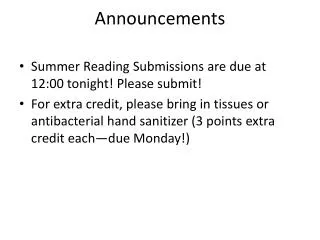
Announcements. Please remove homework, exam answer sheets from your folders. Answer key is posted outside BR 179. Reading for today: pp. 74-100 on cellular organization Reading for Friday: 154-171 on membrane lipids. I. Microscopy (cont’d) E. Electron Microscopy 1. TEM 2. SEM


Announcements
E N D
Presentation Transcript
Announcements • Please remove homework, exam answer sheets from your folders. • Answer key is posted outside BR 179. • Reading for today: pp. 74-100 on cellular organization • Reading for Friday: 154-171 on membrane lipids
I. Microscopy (cont’d) E. Electron Microscopy 1. TEM 2. SEM II. Cellular Organization A. “Prokaryotes” versus Eukaryotes B. Isolation and fractionation of eukaryotic organelles and proteins C. Eukaryotic organelles After reading the text, attending lecture, and reviewing lecture notes, you should be able to: Compare and contrast scanning versus transmission EM. Compare and contrast “prokaryotic” and eukaryotic cells Explain how cells are purified and how their contents are fractionated, including techniques of differential centrifugation and column chromatography. State the primary function of each eukaryotic organelle. Apply your knowledge of these topics to cell biological problems. Outline/Learning Outcomes
Epifluorescence/Confocal light path Conventional epifluorescence Confocal epifluorescence
Comparison of resolving power Light microscopy Electron microscopy Why does electron microscopy have greater resolving power?
Electron Microscopy • How do we get higher resolution than 0.2 μm? • Wavelength () of an electron (at 1,000,000 V) = 0.001 nm (versus 450 nm for LM). • If NA = 0.004 (versus 1.4 for LM), r = 0.61(0.001nm)/0.004 = 0.2 nm • This is 1000-fold better than the best light microscope. Philips CM10 TEM in CMU Biology Microscopy Facility
Scanning EM • Primary electrons hit sample and produce secondary electrons, which are collected and converted to photons on a video screen. • Electron beam scans across sample to produce image. • Large depth of field possible, good for 3-D • Used together with freeze-fracture technique to study membranes. JEOL JSM-840A SEM in CMU Biology Microcopy Facility
Examples of SEM (a) Cultured human neuroblastoma, (b) pollen grain
“Prokaryotes” – a paraphyletic group (doesn’t include all descendants of last common ancestor) DOMAINS
“Prokaryotic” versus Eukaryotic Cells cytoskeleton
Limits on cell size • How large depends on: • Sufficient surface area/volume ratio • Rates of diffusion of molecules • Adequate local concentrations of substrates and enzymes • How small depends on: • Minimal number of substrates, enzymes • Sufficient volume to package the DNA Collisions with other molecules cause diffusion from higher to lower concentration
Isolation of Organelles A. Cell purification by centrifugation, cell culture, etc. B. Cell disruption by grinding, sonication, or osmotic lysis. C. Cell fractionation by differential centrifugation D. Characterization by activity of enzyme markers, EM, etc. Fig. 12A-1
Differential Centrifugation • Particles separate based on different rates of sedimentation due to size, shape, or density • In velocity sedimentation: • 1000 X g, 10 min → nuclear fraction • 20,000 X g, 20 min → mitochondrial fraction • 80,000 X g, 1 hr → microsomal fraction (membranes) • Remaining supernatant: cytosol • 150,000+ X g, 3 hr → macromolecules Fig. 12A-4
Sedimentation Coefficients • Particles with large sedimentation coefficients (S) pellet sooner than those with smaller ones. • Organelles, macromolecules, and viruses can be separated by their density (by density gradient centrifugation) or size: Fig. 12A-2, 12A-3 5S-80S for rRNA
Enzyme Assays for Purity(Don’t memorize) • Plasma membrane: Na/K ATPase • Nucleus: DNA • Golgi: UDP-galactosyl transferase • Mitochondria: monoamine oxidase, cytochrome oxidase, etc. • Lysosomes: acid phosphatase • Peroxisomes: catalase • Endoplasmic reticulum (rough): glucose-6-phosphatase
Three kinds of chromatography • Ion-exchange: separation by charge • Gel-filtration: separation by size • Affinity: separation by specific binding
HPLC • Highest resolution obtained by high-performance liquid chromatography, using tiny spheres and solvent under pressure.
Learning check The purification of a protein usually requires multiple steps and often involves several types of chromatography. A key component of any purification is an assay for the desired protein. The assay can be a band on a gel, a structure in the electron microscope, the ability to bind to another molecule, or an enzymatic activity. The purification of an enzyme is particularly instructive because the assay allows one to quantify the extent of purification at each step. Consider the purification of the enzyme shown below. The total volume, total protein, and total enzyme activity are shown at each step. (A) For each step, calculate the specific activity. (B) Which purification step was most effective? Which step was the least effective? (C) How would S.A. change if carried through further purification steps? How would you know when the enzyme was totally pure? *1 unit of enzyme activity defined as amount converting 1 μmol substrate per min at 25oC.
3.5X 6.5X 3.8X 17X Learning check The purification of a protein usually requires multiple steps and often involves several types of chromatography. A key component of any purification is an assay for the desired protein. The assay can be a band on a gel, a structure in the electron microscope, the ability to bind to another molecule, or an enzymatic activity. The purification of an enzyme is particularly instructive because the assay allows one to quantify the extent of purification at each step. Consider the purification of the enzyme shown below. The total volume, total protein, and total enzyme activity are shown at each step. (A) For each step, calculate the specific activity. (B) Which purification step was most effective? Which step was the least effective? (C) How would S.A. change if carried through further purification steps? How would you know when the enzyme was totally pure?
The Secretory Pathway Endoplasmic Reticulum Transition vesicles Golgi complex Secretory vesicles Plasma membrane